


 Foundational characteristics of cancer include proliferation, angiogenesis, migration, evasion of apoptosis, and cellular immortality. Find key markers for these cellular processes and antibodies to detect them.
 The SUMOplot™ Analysis Program predicts and scores sumoylation sites in your protein. SUMOylation is a post-translational modification involved in various cellular processes, such as nuclear-cytosolic transport, transcriptional regulation, apoptosis, protein stability, response to stress, and progression through the cell cycle.
 The Autophagy Receptor Motif Plotter predicts and scores autophagy receptor binding sites in your protein. Identifying proteins connected to this pathway is critical to understanding the role of autophagy in physiological as well as pathological processes such as development, differentiation, neurodegenerative diseases, stress, infection, and cancer.
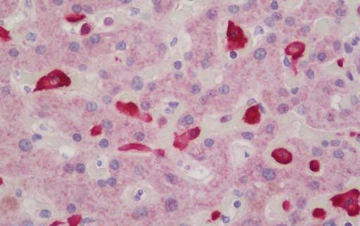
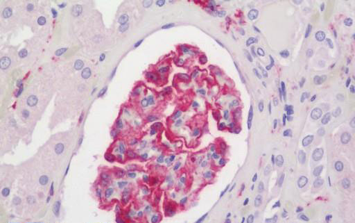
AIF1 / IBA1 Antibody
Rabbit Polyclonal Antibody
- SPECIFICATION
- CITATIONS
- PROTOCOLS
- BACKGROUND

Application
| WB, IHC-P, E |
|---|---|
| Primary Accession | P55008 |
| Other Accession | 199 |
| Reactivity | Human, Mouse, Rat |
| Host | Rabbit |
| Clonality | Polyclonal |
| Calculated MW | 16703 Da |
| Dilution | ELISA (1:10000 - 1:40000), IHC-P (5 µg/ml), WB (1:500 - 1:1000), |
| Gene ID | 199 |
|---|---|
| Other Names | AIF1, AIF-1, Em:AF129756.17, G1, IRT-1, Protein G1, IBA1, IRT1 |
| Reconstitution & Storage | PBS, pH 7.4, 0.02% sodium azide. Store at -20°C for up to one year. |
| Precautions | AIF1 / IBA1 Antibody is for research use only and not for use in diagnostic or therapeutic procedures. |
| Name | AIF1 |
|---|---|
| Synonyms | G1, IBA1 |
| Function | Actin-binding protein that enhances membrane ruffling and RAC activation. Enhances the actin-bundling activity of LCP1. Binds calcium. Plays a role in RAC signaling and in phagocytosis. May play a role in macrophage activation and function. Promotes the proliferation of vascular smooth muscle cells and of T-lymphocytes. Enhances lymphocyte migration. Plays a role in vascular inflammation. |
| Cellular Location | Cytoplasm, cytoskeleton {ECO:0000250|UniProtKB:O70200}. Cell projection, ruffle membrane {ECO:0000250|UniProtKB:O70200}; Peripheral membrane protein {ECO:0000250|UniProtKB:O70200}; Cytoplasmic side {ECO:0000250|UniProtKB:O70200}. Cell projection, phagocytic cup {ECO:0000250|UniProtKB:O70200}. Note=Associated with the actin cytoskeleton at membrane ruffles and at sites of phagocytosis {ECO:0000250|UniProtKB:O70200} |
| Tissue Location | Detected in T-lymphocytes and peripheral blood mononuclear cells. |
| Volume | 50 µl |

Thousands of laboratories across the world have published research that depended on the performance of antibodies from Abcepta to advance their research. Check out links to articles that cite our products in major peer-reviewed journals, organized by research category.
info@abcepta.com, and receive a free "I Love Antibodies" mug.

Provided below are standard protocols that you may find useful for product applications.
Background
Actin-binding protein that enhances membrane ruffling and RAC activation. Enhances the actin-bundling activity of LCP1. Binds calcium. Plays a role in RAC signaling and in phagocytosis. May play a role in macrophage activation and function. Promotes the proliferation of vascular smooth muscle cells and of T- lymphocytes. Enhances lymphocyte migration. Plays a role in vascular inflammation.
References
Albertella M.R.,et al.Hum. Mol. Genet. 3:793-799(1994).
Holzinger I.,et al.Immunogenetics 42:315-322(1995).
Autieri M.V.,et al.Biochem. Biophys. Res. Commun. 228:29-37(1996).
Utans U.,et al.Transplantation 61:1387-1392(1996).
de Baey A.,et al.Genomics 45:591-600(1997).
If you have used an Abcepta product and would like to share how it has performed, please click on the "Submit Review" button and provide the requested information. Our staff will examine and post your review and contact you if needed.
If you have any additional inquiries please email technical services at tech@abcepta.com.
Ordering Information
Other Products
Shipping Information
