


 Foundational characteristics of cancer include proliferation, angiogenesis, migration, evasion of apoptosis, and cellular immortality. Find key markers for these cellular processes and antibodies to detect them.
 The SUMOplot™ Analysis Program predicts and scores sumoylation sites in your protein. SUMOylation is a post-translational modification involved in various cellular processes, such as nuclear-cytosolic transport, transcriptional regulation, apoptosis, protein stability, response to stress, and progression through the cell cycle.
 The Autophagy Receptor Motif Plotter predicts and scores autophagy receptor binding sites in your protein. Identifying proteins connected to this pathway is critical to understanding the role of autophagy in physiological as well as pathological processes such as development, differentiation, neurodegenerative diseases, stress, infection, and cancer.
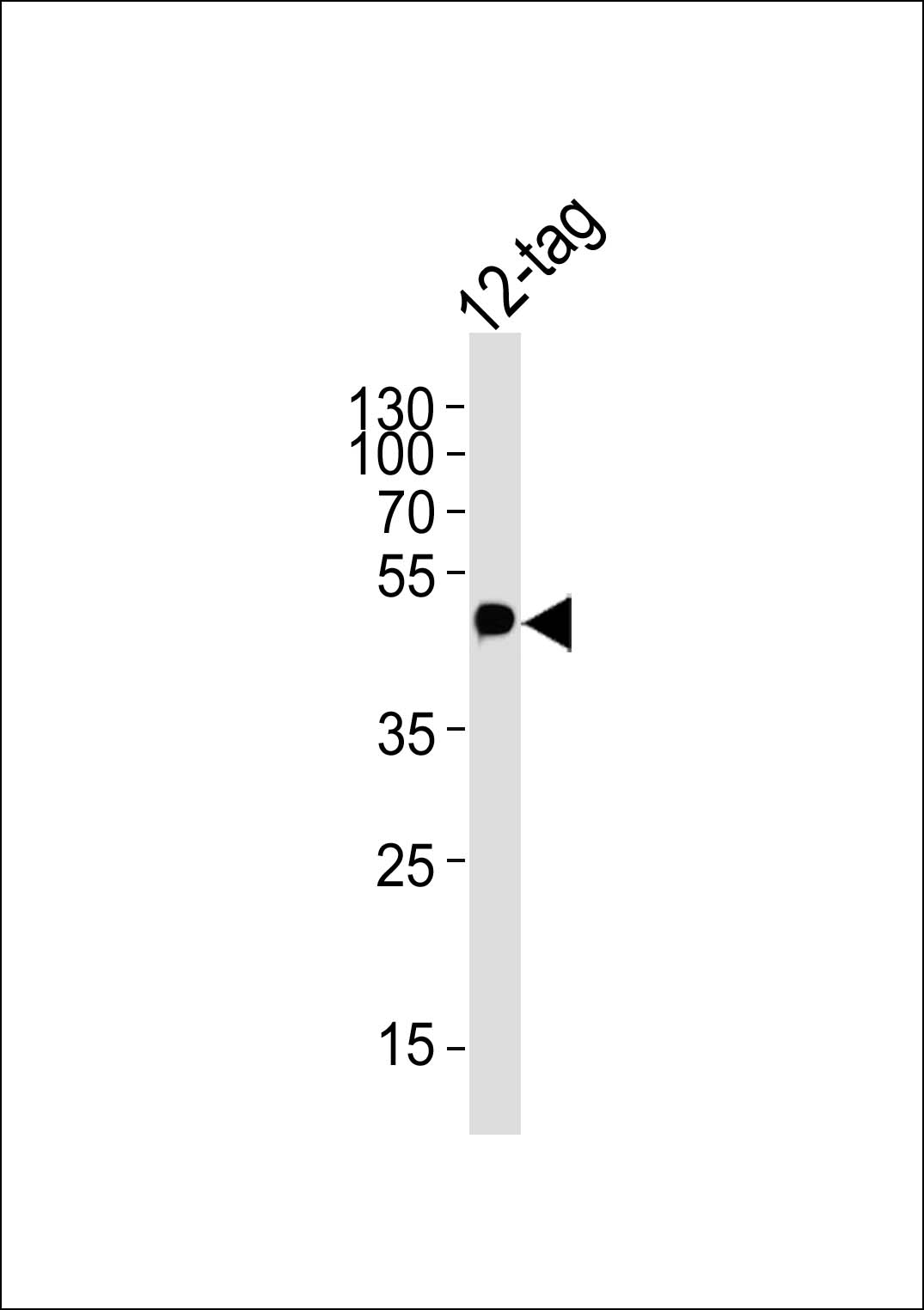
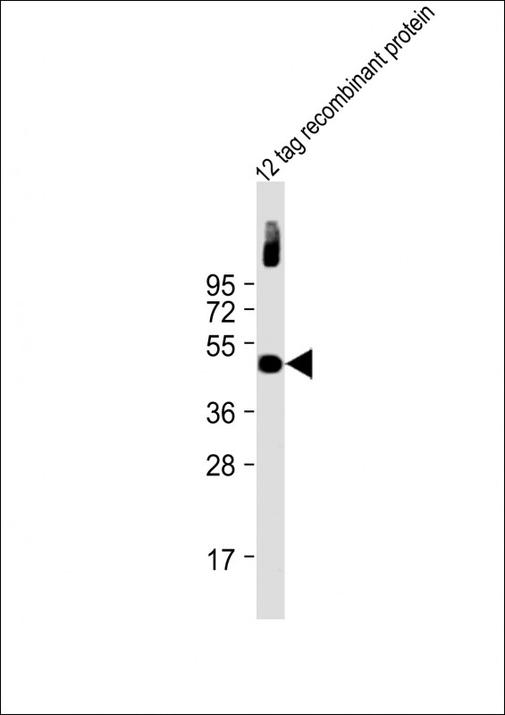
Myc Tag Antibody
Purified Mouse Monoclonal Antibody (Mab)
- SPECIFICATION
- CITATIONS: 18
- PROTOCOLS
- BACKGROUND

Application
| WB, E |
|---|---|
| Host | Mouse |
| Clonality | Monoclonal |
| Isotype | Mouse IgG1 |
| Clone/Animal Names | 9E10 |
| Other Names | Tag from c-Myc protein |
|---|---|
| Target/Specificity | KLH conjugated synthetic peptide encoding c-Myc tag (AEEQKLISEEDLLRKRREQLKHKLE) was used as antigen. |
| Dilution | WB~~1:2000 E~~Use at an assay dependent concentration. |
| Format | Purified monoclonal antibody supplied in PBS with 0.09% (W/V) sodium azide. This antibody is purified through a protein G column, followed by dialysis against PBS. |
| Storage | Maintain refrigerated at 2-8°C for up to 2 weeks. For long term storage store at -20°C in small aliquots to prevent freeze-thaw cycles. |
| Precautions | Myc Tag Antibody is for research use only and not for use in diagnostic or therapeutic procedures. |

Provided below are standard protocols that you may find useful for product applications.
Background
Epitope tags consisting of short sequences recognized by well-characterizated monoclonal antibodies have been widely used in the study of protein expression in various systems. The HA tag (YPYDVPDYA) and Myc Tag (AEEQKLISEEDLLRKRREQLKHKLE), recognized by monoclonal antibody clones 12CA5 and 9E10, respectively, are illustrative examples. Abgent's anti-Myc monoclonal antibody (Clone 9E10) provides a simple solution to detect the expression of a Myc-tagged protein in cells.
References
Kolodziej, PA and Young RA. (1991) Methods Enzymol., 194:508-19. Sells MA and Chernoff J. (1995) Gene, 152:187-9.
If you have used an Abcepta product and would like to share how it has performed, please click on the "Submit Review" button and provide the requested information. Our staff will examine and post your review and contact you if needed.
If you have any additional inquiries please email technical services at tech@abcepta.com.
Ordering Information
Shipping Information
