


 Foundational characteristics of cancer include proliferation, angiogenesis, migration, evasion of apoptosis, and cellular immortality. Find key markers for these cellular processes and antibodies to detect them.
 The SUMOplot™ Analysis Program predicts and scores sumoylation sites in your protein. SUMOylation is a post-translational modification involved in various cellular processes, such as nuclear-cytosolic transport, transcriptional regulation, apoptosis, protein stability, response to stress, and progression through the cell cycle.
 The Autophagy Receptor Motif Plotter predicts and scores autophagy receptor binding sites in your protein. Identifying proteins connected to this pathway is critical to understanding the role of autophagy in physiological as well as pathological processes such as development, differentiation, neurodegenerative diseases, stress, infection, and cancer.
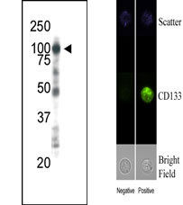
AC133 (CD133) Antibody
Purified Mouse Monoclonal Antibody (Mab)
- SPECIFICATION
- CITATIONS
- PROTOCOLS
- BACKGROUND

Application
| WB, IHC-P, E |
|---|---|
| Primary Accession | O43490 |
| Reactivity | Human |
| Host | Mouse |
| Clonality | Monoclonal |
| Isotype | Mouse IgG1 |
| Clone/Animal Names | 32AT1672 |
| Calculated MW | 97202 Da |
| Gene ID | 8842 |
|---|---|
| Other Names | Prominin-1, Antigen AC133, Prominin-like protein 1, CD133, PROM1, PROML1 |
| Target/Specificity | This monoclonal antibody is generated from mice immunized with a KLH conjugated synthetic peptide selected from the N-terminal region of AC133. |
| Dilution | WB~~1:500~1000 IHC-P~~1:10~50 E~~Use at an assay dependent concentration. |
| Format | Purified polyclonal antibody supplied in PBS with 0.09% (W/V) sodium azide. This antibody is purified through a protein G column, followed by dialysis against PBS. |
| Storage | Maintain refrigerated at 2-8°C for up to 2 weeks. For long term storage store at -20°C in small aliquots to prevent freeze-thaw cycles. |
| Precautions | AC133 (CD133) Antibody is for research use only and not for use in diagnostic or therapeutic procedures. |
| Name | PROM1 |
|---|---|
| Synonyms | PROML1 |
| Function | May play a role in cell differentiation, proliferation and apoptosis (PubMed:24556617). Binds cholesterol in cholesterol- containing plasma membrane microdomains and may play a role in the organization of the apical plasma membrane in epithelial cells. During early retinal development acts as a key regulator of disk morphogenesis. Involved in regulation of MAPK and Akt signaling pathways. In neuroblastoma cells suppresses cell differentiation such as neurite outgrowth in a RET-dependent manner (PubMed:20818439). |
| Cellular Location | Apical cell membrane; Multi-pass membrane protein. Cell projection, microvillus membrane; Multi-pass membrane protein. Cell projection, cilium, photoreceptor outer segment Endoplasmic reticulum. Endoplasmic reticulum-Golgi intermediate compartment. Note=Found in extracellular membrane particles in various body fluids such as cerebrospinal fluid, saliva, seminal fluid and urine |
| Tissue Location | Isoform 1 is selectively expressed on CD34 hematopoietic stem and progenitor cells in adult and fetal bone marrow, fetal liver, cord blood and adult peripheral blood. Isoform 1 is not detected on other blood cells. Isoform 1 is also expressed in a number of non-lymphoid tissues including retina, pancreas, placenta, kidney, liver, lung, brain and heart. Found in saliva within small membrane particles. Isoform 2 is predominantly expressed in fetal liver, skeletal muscle, kidney, and heart as well as adult pancreas, kidney, liver, lung, and placenta. Isoform 2 is highly expressed in fetal liver, low in bone marrow, and barely detectable in peripheral blood Isoform 2 is expressed on hematopoietic stem cells and in epidermal basal cells (at protein level). Expressed in adult retina by rod and cone photoreceptor cells (at protein level) |

Thousands of laboratories across the world have published research that depended on the performance of antibodies from Abcepta to advance their research. Check out links to articles that cite our products in major peer-reviewed journals, organized by research category.
info@abcepta.com, and receive a free "I Love Antibodies" mug.

Provided below are standard protocols that you may find useful for product applications.
Background
This gene encodes a pentaspan transmembrane glycoprotein. The protein localizes to membrane protrusions and is often expressed on adult stem cells, where it is thought to function in maintaining stem cell properties by suppressing differentiation. Mutations in this gene have been shown to result in retinitis pigmentosa and Stargardt disease. Expression of this gene is also associated with several types of cancer. This gene is expressed from at least five alternative promoters that are expressed in a tissue-dependent manner. Multiple transcript variants encoding different isoforms have been found for this gene.
References
Prognostic impact of the expression of putative cancer stem cell markers CD133, CD166, CD44s, EpCAM, and ALDH1 in colorectal cancer. Lugli A, et al. Br J Cancer, 2010 Jul 27. PMID 20606680. Members of the low-density lipoprotein receptor-related proteins provide a differential molecular signature between parental and CD133+ DAOY medulloblastoma cells. Annabi B, et al. Mol Carcinog, 2010 Jul. PMID 20564348. Activation of Akt and MAPK pathways enhances the tumorigenicity of CD133+ primary colon cancer cells. Wang YK, et al. Carcinogenesis, 2010 Aug. PMID 20530554. Demethylation of the CD133 gene is frequently detected in early gastric carcinoma. Hibi K, et al. Anticancer Res, 2010 Apr. PMID 20530428. The stem cell marker CD133 associates with enhanced colony formation and cell motility in colorectal cancer. Elsaba TM, et al. PLoS One, 2010 May 19. PMID 20502714.
If you have used an Abcepta product and would like to share how it has performed, please click on the "Submit Review" button and provide the requested information. Our staff will examine and post your review and contact you if needed.
If you have any additional inquiries please email technical services at tech@abcepta.com.
Ordering Information
Other Products
Shipping Information
