


 Foundational characteristics of cancer include proliferation, angiogenesis, migration, evasion of apoptosis, and cellular immortality. Find key markers for these cellular processes and antibodies to detect them.
 The SUMOplot™ Analysis Program predicts and scores sumoylation sites in your protein. SUMOylation is a post-translational modification involved in various cellular processes, such as nuclear-cytosolic transport, transcriptional regulation, apoptosis, protein stability, response to stress, and progression through the cell cycle.
 The Autophagy Receptor Motif Plotter predicts and scores autophagy receptor binding sites in your protein. Identifying proteins connected to this pathway is critical to understanding the role of autophagy in physiological as well as pathological processes such as development, differentiation, neurodegenerative diseases, stress, infection, and cancer.
RET Antibody (Ascites)
Mouse Monoclonal Antibody (Mab)
- SPECIFICATION
- CITATIONS: 1
- PROTOCOLS
- BACKGROUND

Application
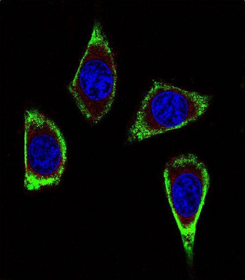
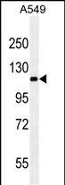
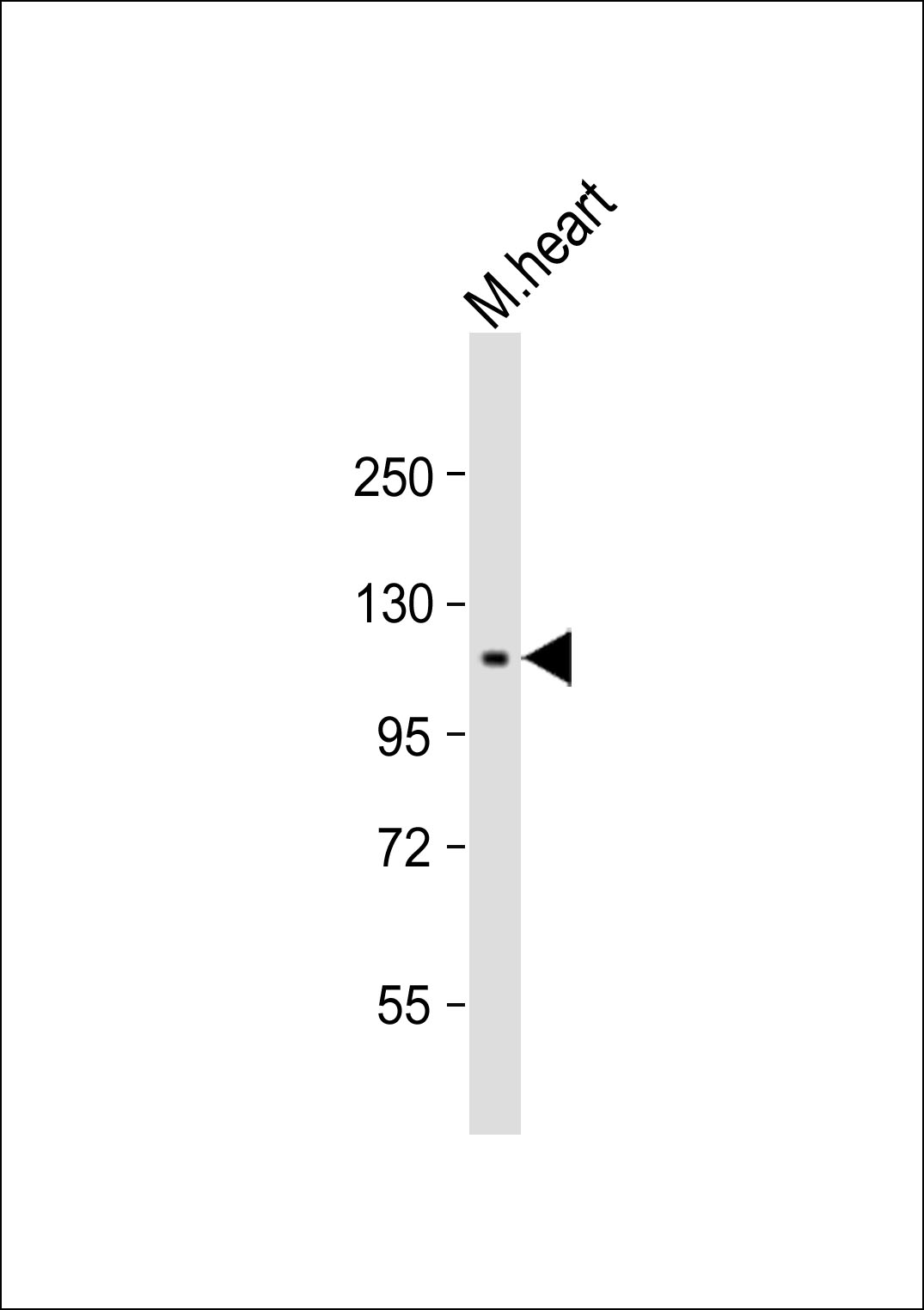
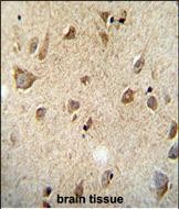
| IF, IHC-P, WB, E |
|---|---|
| Primary Accession | P07949 |
| Other Accession | NP_065681.1, NP_066124.1 |
| Reactivity | Human |
| Host | Mouse |
| Clonality | Monoclonal |
| Isotype | IgM,K |
| Clone/Animal Names | 188CT11.2.3 |
| Calculated MW | 124319 Da |
| Gene ID | 5979 |
|---|---|
| Other Names | Proto-oncogene tyrosine-protein kinase receptor Ret, Cadherin family member 12, Proto-oncogene c-Ret, Soluble RET kinase fragment, Extracellular cell-membrane anchored RET cadherin 120 kDa fragment, RET, CDHF12, CDHR16, PTC, RET51 |
| Target/Specificity | This RET Monoclonal antibody was raised using purified His-tagged recombinant human RET. |
| Dilution | IF~~1:10~50 IHC-P~~1:50~100 WB~~1:1000 E~~Use at an assay dependent concentration. |
| Format | Mouse monoclonal antibody supplied in crude ascites with 0.09% (W/V) sodium azide. |
| Storage | Maintain refrigerated at 2-8°C for up to 2 weeks. For long term storage store at -20°C in small aliquots to prevent freeze-thaw cycles. |
| Precautions | RET Antibody (Ascites) is for research use only and not for use in diagnostic or therapeutic procedures. |
| Name | RET {ECO:0000303|PubMed:2660074, ECO:0000312|HGNC:HGNC:9967} |
|---|---|
| Function | Receptor tyrosine-protein kinase involved in numerous cellular mechanisms including cell proliferation, neuronal navigation, cell migration, and cell differentiation in response to glia cell line- derived growth family factors (GDNF, NRTN, ARTN, PSPN and GDF15) (PubMed:20064382, PubMed:20616503, PubMed:20702524, PubMed:21357690, PubMed:21454698, PubMed:24560924, PubMed:28846097, PubMed:28846099, PubMed:28953886, PubMed:31118272). In contrast to most receptor tyrosine kinases, RET requires not only its cognate ligands but also coreceptors, for activation (PubMed:21994944, PubMed:23333276, PubMed:28846097, PubMed:28846099, PubMed:28953886). GDNF ligands (GDNF, NRTN, ARTN, PSPN and GDF15) first bind their corresponding GDNFR coreceptors (GFRA1, GFRA2, GFRA3, GFRA4 and GFRAL, respectively), triggering RET autophosphorylation and activation, leading to activation of downstream signaling pathways, including the MAPK- and AKT-signaling pathways (PubMed:21994944, PubMed:23333276, PubMed:24560924, PubMed:25242331, PubMed:28846097, PubMed:28846099, PubMed:28953886). Acts as a dependence receptor via the GDNF-GFRA1 signaling: in the presence of the ligand GDNF in somatotrophs within pituitary, promotes survival and down regulates growth hormone (GH) production, but triggers apoptosis in absence of GDNF (PubMed:20616503, PubMed:21994944). Required for the molecular mechanisms orchestration during intestine organogenesis via the ARTN-GFRA3 signaling: involved in the development of enteric nervous system and renal organogenesis during embryonic life, and promotes the formation of Peyer's patch-like structures, a major component of the gut-associated lymphoid tissue (By similarity). Mediates, through interaction with GDF15-receptor GFRAL, GDF15-induced cell-signaling in the brainstem which triggers an aversive response, characterized by nausea, vomiting, and/or loss of appetite in response to various stresses (PubMed:28846097, PubMed:28846099, PubMed:28953886). Modulates cell adhesion via its cleavage by caspase in sympathetic neurons and mediates cell migration in an integrin (e.g. ITGB1 and ITGB3)-dependent manner (PubMed:20702524, PubMed:21357690). Also active in the absence of ligand, triggering apoptosis through a mechanism that requires receptor intracellular caspase cleavage (PubMed:21357690). Triggers the differentiation of rapidly adapting (RA) mechanoreceptors (PubMed:20064382). Involved in the development of the neural crest (By similarity). Regulates nociceptor survival and size (By similarity). Phosphorylates PTK2/FAK1 (PubMed:21454698). |
| Cellular Location | Cell membrane; Single-pass type I membrane protein. Endosome membrane; Single-pass type I membrane protein Note=Predominantly located on the plasma membrane (PubMed:23333276, PubMed:9575150). In the presence of SORL1 and GFRA1, directed to endosomes (PubMed:23333276). |

Provided below are standard protocols that you may find useful for product applications.
Background
This gene, a member of the cadherin superfamily, encodes one of the receptor tyrosine kinases, which are cell-surface molecules that transduce signals for cell growth and differentiation. This gene plays a crucial role in neural crest development, and it can undergo oncogenic activation in vivo and in vitro by cytogenetic rearrangement. Mutations in this gene are associated with the disorders multiple endocrine neoplasia, type IIA, multiple endocrine neoplasia, type IIB, Hirschsprung disease, and medullary thyroid carcinoma. Two transcript variants encoding different isoforms have been found for this gene. Additional transcript variants have been described but their biological validity has not been confirmed.
References
Siqueira, D.R., et al. Endocr. Relat. Cancer 17(4):953-963(2010)
Gockel, H.R., et al. Hum. Genet. 128(4):353-364(2010)
Kim, H.K., et al. Anticancer Res. 30(9):3621-3627(2010)
Pacini, F., et al. Clin Oncol (R Coll Radiol) 22(6):475-485(2010)
Jugessur, A., et al. PLoS ONE 5 (7), E11493 (2010) :
If you have used an Abcepta product and would like to share how it has performed, please click on the "Submit Review" button and provide the requested information. Our staff will examine and post your review and contact you if needed.
If you have any additional inquiries please email technical services at tech@abcepta.com.
Ordering Information
Other Products
Shipping Information
