


 Foundational characteristics of cancer include proliferation, angiogenesis, migration, evasion of apoptosis, and cellular immortality. Find key markers for these cellular processes and antibodies to detect them.
 The SUMOplot™ Analysis Program predicts and scores sumoylation sites in your protein. SUMOylation is a post-translational modification involved in various cellular processes, such as nuclear-cytosolic transport, transcriptional regulation, apoptosis, protein stability, response to stress, and progression through the cell cycle.
 The Autophagy Receptor Motif Plotter predicts and scores autophagy receptor binding sites in your protein. Identifying proteins connected to this pathway is critical to understanding the role of autophagy in physiological as well as pathological processes such as development, differentiation, neurodegenerative diseases, stress, infection, and cancer.
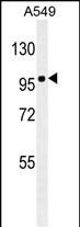
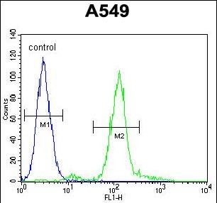
ZEB1 antibody ( Ascites)
Mouse Monoclonal Antibody (Mab)
- SPECIFICATION
- CITATIONS: 2
- PROTOCOLS
- BACKGROUND

Application
| FC, WB, E |
|---|---|
| Primary Accession | P37275 |
| Reactivity | Human |
| Host | Mouse |
| Clonality | Monoclonal |
| Isotype | IgG1,K |
| Clone/Animal Names | 213CT14.1.5 |
| Calculated MW | 124074 Da |
| Antigen Region | 69-97 aa |
| Gene ID | 6935 |
|---|---|
| Other Names | Zinc finger E-box-binding homeobox 1, NIL-2-A zinc finger protein, Negative regulator of IL2, Transcription factor 8, TCF-8, ZEB1, AREB6, TCF8 |
| Target/Specificity | This ZEB1 antibody is generated from mice immunized with a KLH conjugated synthetic peptide between 69-97 amino acids from human ZEB1. |
| Dilution | FC~~1:10~50 WB~~1:500~16000 E~~Use at an assay dependent concentration. |
| Format | Mouse monoclonal antibody supplied in crude ascites with 0.09% (W/V) sodium azide. |
| Storage | Maintain refrigerated at 2-8°C for up to 2 weeks. For long term storage store at -20°C in small aliquots to prevent freeze-thaw cycles. |
| Precautions | ZEB1 antibody ( Ascites) is for research use only and not for use in diagnostic or therapeutic procedures. |
| Name | ZEB1 (HGNC:11642) |
|---|---|
| Function | Acts as a transcriptional repressor. Inhibits interleukin-2 (IL-2) gene expression. Enhances or represses the promoter activity of the ATP1A1 gene depending on the quantity of cDNA and on the cell type. Represses E-cadherin promoter and induces an epithelial-mesenchymal transition (EMT) by recruiting SMARCA4/BRG1. Represses BCL6 transcription in the presence of the corepressor CTBP1. Positively regulates neuronal differentiation. Represses RCOR1 transcription activation during neurogenesis. Represses transcription by binding to the E box (5'-CANNTG-3'). In the absence of TGFB1, acts as a repressor of COL1A2 transcription via binding to the E-box in the upstream enhancer region (By similarity). |
| Cellular Location | Nucleus |
| Tissue Location | Colocalizes with SMARCA4/BRG1 in E-cadherin- negative cells from established lines, and stroma of normal colon as well as in de-differentiated epithelial cells at the invasion front of colorectal carcinomas (at protein level). Expressed in heart and skeletal muscle, but not in liver, spleen, or pancreas |

Provided below are standard protocols that you may find useful for product applications.
Background
This gene encodes a zinc finger transcription factor. The encoded protein likely plays a role in transcriptional repression of interleukin 2. Mutations in this gene have been associated with posterior polymorphous corneal dystrophy-3 and late-onset Fuchs endothelial corneal dystrophy. Alternatively spliced transcript variants encoding different isoforms have been described.
References
Drake, J.M., et al. J. Biol. Chem. 285(44):33940-33948(2010) Takeyama, Y., et al. Cancer Lett. 296(2):216-224(2010) Nakahata, S., et al. Oncogene 29(29):4157-4169(2010) Mehta, J.S., et al. Invest. Ophthalmol. Vis. Sci. 49(1):184-188(2008) Manavella, P.A., et al. Biochem. Biophys. Res. Commun. 360(3):621-626(2007)
If you have used an Abcepta product and would like to share how it has performed, please click on the "Submit Review" button and provide the requested information. Our staff will examine and post your review and contact you if needed.
If you have any additional inquiries please email technical services at tech@abcepta.com.
Ordering Information
Other Products
Shipping Information
