


 Foundational characteristics of cancer include proliferation, angiogenesis, migration, evasion of apoptosis, and cellular immortality. Find key markers for these cellular processes and antibodies to detect them.
 The SUMOplot™ Analysis Program predicts and scores sumoylation sites in your protein. SUMOylation is a post-translational modification involved in various cellular processes, such as nuclear-cytosolic transport, transcriptional regulation, apoptosis, protein stability, response to stress, and progression through the cell cycle.
 The Autophagy Receptor Motif Plotter predicts and scores autophagy receptor binding sites in your protein. Identifying proteins connected to this pathway is critical to understanding the role of autophagy in physiological as well as pathological processes such as development, differentiation, neurodegenerative diseases, stress, infection, and cancer.
TGFB2 Antibody
Mouse Monoclonal Antibody (Mab)
- SPECIFICATION
- CITATIONS
- PROTOCOLS
- BACKGROUND

Application
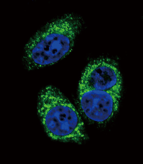
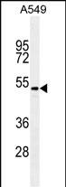
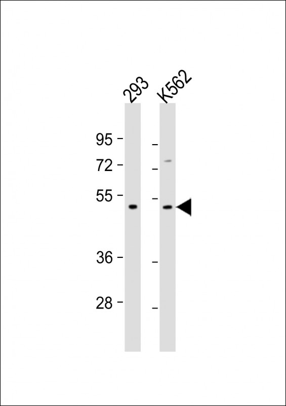
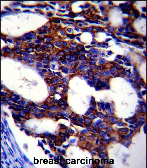
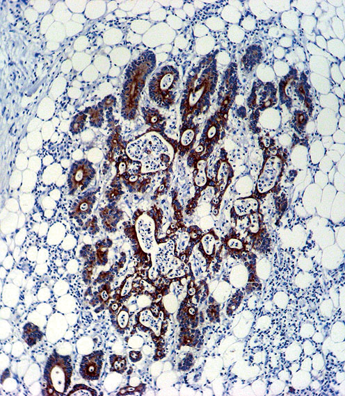
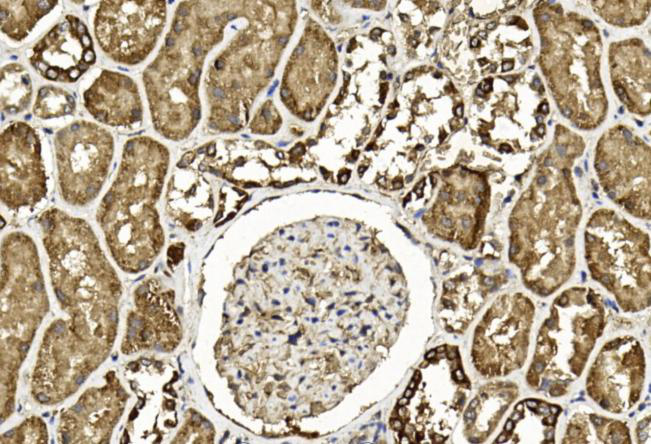
| WB, IF, IHC-P, E |
|---|---|
| Primary Accession | P61812 |
| Other Accession | NP_001129071.1, NP_003229.1 |
| Reactivity | Human |
| Host | Mouse |
| Clonality | Monoclonal |
| Isotype | IgG1,K |
| Clone/Animal Names | 220ct16.4.3.1 |
| Calculated MW | 47748 Da |
| Gene ID | 7042 |
|---|---|
| Other Names | Transforming growth factor beta-2, TGF-beta-2, BSC-1 cell growth inhibitor, Cetermin, Glioblastoma-derived T-cell suppressor factor, G-TSF, Polyergin, Latency-associated peptide, LAP, TGFB2 |
| Target/Specificity | This TGFB2 monoclonal antibody is generated from mouse immunized with TGFB2 recombinant protein. |
| Dilution | WB~~1:500-1:1000 IF~~1:10~50 IHC-P~~1:10~50 E~~Use at an assay dependent concentration. |
| Format | Purified monoclonal antibody supplied in PBS with 0.09% (W/V) sodium azide. This antibody is purified through a protein G column, followed by dialysis against PBS. |
| Storage | Maintain refrigerated at 2-8°C for up to 2 weeks. For long term storage store at -20°C in small aliquots to prevent freeze-thaw cycles. |
| Precautions | TGFB2 Antibody is for research use only and not for use in diagnostic or therapeutic procedures. |
| Name | TGFB2 |
|---|---|
| Function | [Transforming growth factor beta-2 proprotein]: Precursor of the Latency-associated peptide (LAP) and Transforming growth factor beta-2 (TGF-beta-2) chains, which constitute the regulatory and active subunit of TGF-beta-2, respectively. |
| Cellular Location | [Latency-associated peptide]: Secreted, extracellular space, extracellular matrix {ECO:0000250|UniProtKB:P01137} |

Thousands of laboratories across the world have published research that depended on the performance of antibodies from Abcepta to advance their research. Check out links to articles that cite our products in major peer-reviewed journals, organized by research category.
info@abcepta.com, and receive a free "I Love Antibodies" mug.

Provided below are standard protocols that you may find useful for product applications.
Background
This gene encodes a member of the transforming growth factor beta (TGFB) family of cytokines, which are multifunctional peptides that regulate proliferation, differentiation, adhesion, migration, and other functions in many cell types by transducing their signal through combinations of transmembrane type I and type II receptors (TGFBR1 and TGFBR2) and their downstream effectors, the SMAD proteins. Disruption of the TGFB/SMAD pathway has been implicated in a variety of human cancers. The encoded protein is secreted and has suppressive effects of interleukin-2 dependent T-cell growth. Translocation t(1;7)(q41;p21) between this gene and HDAC9 is associated with Peters' anomaly, a congenital defect of the anterior chamber of the eye. The knockout mice lacking this gene show perinatal mortality and a wide range of developmental, including cardiac, defects. Alternatively spliced transcript variants encoding different isoforms have been identified.
References
Nalpas, B., et al. Gut 59(8):1120-1126(2010)
Bailey, S.D., et al. Diabetes Care (2010) In press :
Jugessur, A., et al. PLoS ONE 5 (7), E11493 (2010) :
Johnatty, S.E., et al. PLoS Genet. 6 (7), E1001016 (2010) :
Sambo, M.R., et al. PLoS ONE 5 (6), E11141 (2010) :
If you have used an Abcepta product and would like to share how it has performed, please click on the "Submit Review" button and provide the requested information. Our staff will examine and post your review and contact you if needed.
If you have any additional inquiries please email technical services at tech@abcepta.com.
Ordering Information
Other Products
Shipping Information
