


 Foundational characteristics of cancer include proliferation, angiogenesis, migration, evasion of apoptosis, and cellular immortality. Find key markers for these cellular processes and antibodies to detect them.
 The SUMOplot™ Analysis Program predicts and scores sumoylation sites in your protein. SUMOylation is a post-translational modification involved in various cellular processes, such as nuclear-cytosolic transport, transcriptional regulation, apoptosis, protein stability, response to stress, and progression through the cell cycle.
 The Autophagy Receptor Motif Plotter predicts and scores autophagy receptor binding sites in your protein. Identifying proteins connected to this pathway is critical to understanding the role of autophagy in physiological as well as pathological processes such as development, differentiation, neurodegenerative diseases, stress, infection, and cancer.
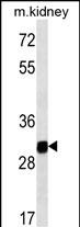
GNPDA1 Antibody
Mouse Monoclonal Antibody (Mab)
- SPECIFICATION
- CITATIONS
- PROTOCOLS
- BACKGROUND

Application
| WB, E |
|---|---|
| Primary Accession | P46926 |
| Other Accession | NP_005462.1 |
| Reactivity | Mouse |
| Host | Mouse |
| Clonality | Monoclonal |
| Isotype | IgG1,k |
| Clone/Animal Names | 237CT2.5.2 |
| Calculated MW | 32669 Da |
| Gene ID | 10007 |
|---|---|
| Other Names | Glucosamine-6-phosphate isomerase 1, Glucosamine-6-phosphate deaminase 1, GNPDA 1, GlcN6P deaminase 1, Oscillin, GNPDA1, GNPI, HLN, KIAA0060 |
| Target/Specificity | This GNPDA1 monoclonal antibody is generated from mouse immunized with GNPDA1 recombinant protein. |
| Dilution | WB~~1:100~1000 E~~Use at an assay dependent concentration. |
| Format | Purified monoclonal antibody supplied in PBS with 0.09% (W/V) sodium azide. This antibody is purified through a protein G column, followed by dialysis against PBS. |
| Storage | Maintain refrigerated at 2-8°C for up to 2 weeks. For long term storage store at -20°C in small aliquots to prevent freeze-thaw cycles. |
| Precautions | GNPDA1 Antibody is for research use only and not for use in diagnostic or therapeutic procedures. |
| Name | GNPDA1 {ECO:0000303|PubMed:26887390, ECO:0000312|HGNC:HGNC:4417} |
|---|---|
| Function | Catalyzes the reversible conversion of alpha-D-glucosamine 6- phosphate (GlcN-6P) into beta-D-fructose 6-phosphate (Fru-6P) and ammonium ion, a regulatory reaction step in de novo uridine diphosphate-N-acetyl-alpha-D-glucosamine (UDP-GlcNAc) biosynthesis via hexosamine pathway. Deamination is coupled to aldo-keto isomerization mediating the metabolic flux from UDP-GlcNAc toward Fru-6P. At high ammonium level can drive amination and isomerization of Fru-6P toward hexosamines and UDP-GlcNAc synthesis (PubMed:21807125, PubMed:26887390). Has a role in fine tuning the metabolic fluctuations of cytosolic UDP-GlcNAc and their effects on hyaluronan synthesis that occur during tissue remodeling (PubMed:26887390). Seems to trigger calcium oscillations in mammalian eggs. These oscillations serve as the essential trigger for egg activation and early development of the embryo (By similarity). |
| Cellular Location | Cytoplasm {ECO:0000250|UniProtKB:O88958}. |

Thousands of laboratories across the world have published research that depended on the performance of antibodies from Abcepta to advance their research. Check out links to articles that cite our products in major peer-reviewed journals, organized by research category.
info@abcepta.com, and receive a free "I Love Antibodies" mug.

Provided below are standard protocols that you may find useful for product applications.
Background
Glucosamine-6-phosphate deaminase (EC 3.5.99.6) is an allosteric enzyme that catalyzes the reversible conversion of D-glucosamine-6-phosphate into D-fructose-6-phosphate and ammonium (Arreola et al., 2003 [PubMed 12965206]).
References
Lamesch, P., et al. Genomics 89(3):307-315(2007) Arreola, R., et al. FEBS Lett. 551 (1-3), 63-70 (2003) : Zhang, J., et al. J. Cell. Biochem. 88(5):932-940(2003) Nakamura, Y., et al. Genomics 68(2):179-186(2000) Shevchenko, V., et al. Gene 216(1):31-38(1998)
If you have used an Abcepta product and would like to share how it has performed, please click on the "Submit Review" button and provide the requested information. Our staff will examine and post your review and contact you if needed.
If you have any additional inquiries please email technical services at tech@abcepta.com.
Ordering Information
Other Products
Shipping Information
