


 Foundational characteristics of cancer include proliferation, angiogenesis, migration, evasion of apoptosis, and cellular immortality. Find key markers for these cellular processes and antibodies to detect them.
 The SUMOplot™ Analysis Program predicts and scores sumoylation sites in your protein. SUMOylation is a post-translational modification involved in various cellular processes, such as nuclear-cytosolic transport, transcriptional regulation, apoptosis, protein stability, response to stress, and progression through the cell cycle.
 The Autophagy Receptor Motif Plotter predicts and scores autophagy receptor binding sites in your protein. Identifying proteins connected to this pathway is critical to understanding the role of autophagy in physiological as well as pathological processes such as development, differentiation, neurodegenerative diseases, stress, infection, and cancer.
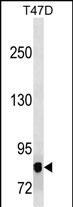
MAP3K12 Antibody
Mouse Monoclonal Antibody (Mab)
- SPECIFICATION
- CITATIONS
- PROTOCOLS
- BACKGROUND

Application
| WB, E |
|---|---|
| Primary Accession | Q12852 |
| Other Accession | NP_001180440.1 |
| Reactivity | Human |
| Host | Mouse |
| Clonality | Monoclonal |
| Isotype | IgG1 |
| Clone/Animal Names | 473CT23.12.5 |
| Calculated MW | 93219 Da |
| Antigen Region | 828-859 aa |
| Gene ID | 7786 |
|---|---|
| Other Names | Mitogen-activated protein kinase kinase kinase 12, Dual leucine zipper bearing kinase, DLK, Leucine-zipper protein kinase, ZPK, MAPK-upstream kinase, MUK, Mixed lineage kinase, MAP3K12, ZPK |
| Target/Specificity | This MAP3K12 antibody is generated from mice immunized with a KLH conjugated synthetic peptide between 828-859 amino acids from human MAP3K12. |
| Dilution | WB~~1:500~1000 E~~Use at an assay dependent concentration. |
| Format | Purified polyclonal antibody supplied in PBS with 0.09% (W/V) sodium azide. This antibody is prepared by Saturated Ammonium Sulfate (SAS) precipitation followed by dialysis against PBS. |
| Storage | Maintain refrigerated at 2-8°C for up to 2 weeks. For long term storage store at -20°C in small aliquots to prevent freeze-thaw cycles. |
| Precautions | MAP3K12 Antibody is for research use only and not for use in diagnostic or therapeutic procedures. |
| Name | MAP3K12 |
|---|---|
| Synonyms | ZPK |
| Function | Part of a non-canonical MAPK signaling pathway (PubMed:28111074). Activated by APOE, enhances the AP-1-mediated transcription of APP, via a MAP kinase signal transduction pathway composed of MAP2K7 and MAPK1/ERK2 and MAPK3/ERK1 (PubMed:28111074). May be an activator of the JNK/SAPK pathway. |
| Cellular Location | Cytoplasm {ECO:0000250|UniProtKB:Q60700}. Cell membrane {ECO:0000250|UniProtKB:Q60700}. Note=Behaves essentially as an integral membrane protein. {ECO:0000250|UniProtKB:Q60700} |
| Tissue Location | Highly expressed in brain and kidney. |

Thousands of laboratories across the world have published research that depended on the performance of antibodies from Abcepta to advance their research. Check out links to articles that cite our products in major peer-reviewed journals, organized by research category.
info@abcepta.com, and receive a free "I Love Antibodies" mug.

Provided below are standard protocols that you may find useful for product applications.
Background
This gene encodes a member of the serine/threonine protein kinase family. This kinase contains a leucine-zipper domain and is predominately expressed in neuronal cells. The phosphorylation state of this kinase in synaptic terminals was shown to be regulated by membrane depolarization via calcineurin. This kinase forms heterodimers with leucine zipper containing transcription factors, such as cAMP responsive element binding protein (CREB) and MYC, and thus may play a regulatory role in PKA or retinoic acid induced neuronal differentiation. Alternatively spliced transcript variants encoding different proteins have been described.[provided by RefSeq].
References
Clague, J., et al. Mol. Carcinog. 49(2):183-189(2010)
Robitaille, H., et al. J. Invest. Dermatol. 130(1):74-85(2010)
Landa, I., et al. PLoS Genet. 5 (9), E1000637 (2009) :
Daviau, A., et al. Cell. Signal. 21(4):577-587(2009)
Mukhopadhyay, R., et al. Mol. Cell 32(3):371-382(2008)
If you have used an Abcepta product and would like to share how it has performed, please click on the "Submit Review" button and provide the requested information. Our staff will examine and post your review and contact you if needed.
If you have any additional inquiries please email technical services at tech@abcepta.com.
Ordering Information
Other Products
Shipping Information
