


 Foundational characteristics of cancer include proliferation, angiogenesis, migration, evasion of apoptosis, and cellular immortality. Find key markers for these cellular processes and antibodies to detect them.
 The SUMOplot™ Analysis Program predicts and scores sumoylation sites in your protein. SUMOylation is a post-translational modification involved in various cellular processes, such as nuclear-cytosolic transport, transcriptional regulation, apoptosis, protein stability, response to stress, and progression through the cell cycle.
 The Autophagy Receptor Motif Plotter predicts and scores autophagy receptor binding sites in your protein. Identifying proteins connected to this pathway is critical to understanding the role of autophagy in physiological as well as pathological processes such as development, differentiation, neurodegenerative diseases, stress, infection, and cancer.
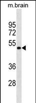
GNAS Antibody (Ascites)
Mouse Monoclonal Antibody (Mab)
- SPECIFICATION
- CITATIONS
- PROTOCOLS
- BACKGROUND

Application
| WB, E |
|---|---|
| Primary Accession | Q5FWY2 |
| Other Accession | P29797, Q8R4A8, P63095, P63094, P63092, P04896, Q63803, Q6R0H7, Q5JWF2 |
| Reactivity | Mouse |
| Predicted | Human, Rat, Bovine, Hamster, Pig |
| Host | Mouse |
| Clonality | Monoclonal |
| Isotype | IgM |
| Clone/Animal Names | 559CT 16.1.3 |
| Calculated MW | 44250 Da |
| Antigen Region | 287-315 aa |
| Gene ID | 2778 |
|---|---|
| Other Names | GNAS complex locus;GNAS; |
| Target/Specificity | This GNAS antibody is generated from mice immunized with a KLH conjugated synthetic peptide between 287-315 amino acids from human GNAS. |
| Dilution | WB~~1:300 E~~Use at an assay dependent concentration. |
| Format | Mouse monoclonal antibody supplied in crude ascites with 0.09% (W/V) sodium azide. |
| Storage | Maintain refrigerated at 2-8°C for up to 2 weeks. For long term storage store at -20°C in small aliquots to prevent freeze-thaw cycles. |
| Precautions | GNAS Antibody (Ascites) is for research use only and not for use in diagnostic or therapeutic procedures. |
| Name | GNAS {ECO:0000313|EMBL:AAH89157.2} |
|---|---|
| Function | Guanine nucleotide-binding protein (G protein) involved as transducer in olfactory signal transduction controlled by G protein- coupled receptors (GPCRs). Contains the guanine nucleotide binding site and alternates between an active, GTP-bound state and an inactive, GDP- bound state. Signaling by an activated GPCR promotes GDP release and GTP binding. The alpha subunit has a low GTPase activity that converts bound GTP to GDP, thereby terminating the signal. Both GDP release and GTP hydrolysis are modulated by numerous regulatory proteins. GNAL/G(olf) alpha specifically mediates olfactory signal transduction within the olfactory neuroepithelium and the basal ganglia following GPCRs activation. Acts by promoting the specific activation of adenylyl cyclase ADCY3, resulting in increased levels of the signaling molecule cAMP. |
| Cellular Location | Cell membrane {ECO:0000256|ARBA:ARBA00004193}; Lipid-anchor {ECO:0000256|ARBA:ARBA00004193} |

Thousands of laboratories across the world have published research that depended on the performance of antibodies from Abcepta to advance their research. Check out links to articles that cite our products in major peer-reviewed journals, organized by research category.
info@abcepta.com, and receive a free "I Love Antibodies" mug.

Provided below are standard protocols that you may find useful for product applications.
Background
Guanine nucleotide-binding proteins (G proteins) are involved as modulators or transducers in various transmembrane signaling systems. The Gs protein is involved in hormonal regulation of adenylate cyclase: it activates the cyclase in response to beta-adrenergic stimuli. Alternative splicing of downstream exons of the GNAS gene is observed, which results in different forms of the stimulatory G protein alpha subunit, a key element of the classical signal transduction pathway linking receptor-ligand interactions with the activation of adenylyl cyclase and a variety of cellular reponses. Multiple transcript variants have been found for this gene, but the full-length nature and/or biological validity of some variants have not been determined. Mutations in this gene result in pseudohypoparathyroidism type 1a, pseudohypoparathyroidism type 1b, Albright hereditary osteodystrophy, pseudopseudohypoparathyroidism, McCune-Albright syndrome, progressive osseus heteroplasia, polyostotic fibrous dysplasia of bone, and some pituitary tumors.
If you have used an Abcepta product and would like to share how it has performed, please click on the "Submit Review" button and provide the requested information. Our staff will examine and post your review and contact you if needed.
If you have any additional inquiries please email technical services at tech@abcepta.com.
Ordering Information
Other Products
Shipping Information
