


 Foundational characteristics of cancer include proliferation, angiogenesis, migration, evasion of apoptosis, and cellular immortality. Find key markers for these cellular processes and antibodies to detect them.
 The SUMOplot™ Analysis Program predicts and scores sumoylation sites in your protein. SUMOylation is a post-translational modification involved in various cellular processes, such as nuclear-cytosolic transport, transcriptional regulation, apoptosis, protein stability, response to stress, and progression through the cell cycle.
 The Autophagy Receptor Motif Plotter predicts and scores autophagy receptor binding sites in your protein. Identifying proteins connected to this pathway is critical to understanding the role of autophagy in physiological as well as pathological processes such as development, differentiation, neurodegenerative diseases, stress, infection, and cancer.
PIp1 Antibody (Center)
Mouse Monoclonal Antibody (Mab)
- SPECIFICATION
- CITATIONS
- PROTOCOLS
- BACKGROUND

Application
| WB, E |
|---|---|
| Primary Accession | P60201 |
| Other Accession | P60202, P60203, P04116, P23289, Q8HXW7, Q712P7, P47789 |
| Reactivity | Human, Mouse, Rat |
| Predicted | Bovine, Chicken, Monkey, Pig, Rabbit |
| Host | Mouse |
| Clonality | Monoclonal |
| Isotype | IgM |
| Clone/Animal Names | 538CT16.5.4 |
| Calculated MW | 30,077 Da |
| Antigen Region | 248-277 aa |
| Gene ID | 5354 |
|---|---|
| Other Names | Myelin proteolipid protein, PLP, Lipophilin, Plp1, Plp |
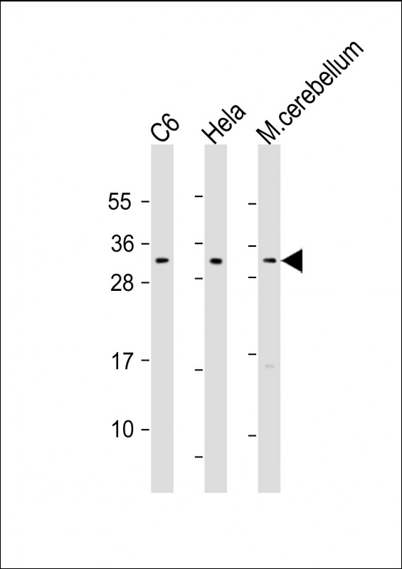
| Target/Specificity | This PIp1 antibody is generated from mice immunized with a KLH conjugated synthetic peptide between 248-277 amino acids from the central region of human PIp1. |
| Dilution | WB~~1:1000 E~~Use at an assay dependent concentration. |
| Format | Purified monoclonal antibody supplied in PBS with 0.09% (W/V) sodium azide. This antibody is prepared by Euglobin precipitation followed by dialysis against PBS. |
| Storage | Maintain refrigerated at 2-8°C for up to 2 weeks. For long term storage store at -20°C in small aliquots to prevent freeze-thaw cycles. |
| Precautions | PIp1 Antibody (Center) is for research use only and not for use in diagnostic or therapeutic procedures. |
| Name | PLP1 |
|---|---|
| Synonyms | PLP |
| Function | This is the major myelin protein from the central nervous system. It plays an important role in the formation or maintenance of the multilamellar structure of myelin. |
| Cellular Location | Cell membrane; Multi-pass membrane protein. Myelin membrane. Note=Colocalizes with SIRT2 in internodal regions, at paranodal axoglial junction and Schmidt-Lanterman incisures of myelin sheat. |

Thousands of laboratories across the world have published research that depended on the performance of antibodies from Abcepta to advance their research. Check out links to articles that cite our products in major peer-reviewed journals, organized by research category.
info@abcepta.com, and receive a free "I Love Antibodies" mug.

Provided below are standard protocols that you may find useful for product applications.
Background
This is the major myelin protein from the central nervous system. It plays an important role in the formation or maintenance of the multilamellar structure of myelin.
References
Mayer, C.A., et al. Respir Physiol Neurobiol 169(3):303-314(2009)
Wang, E., et al. J. Cell. Biochem. 97(5):999-1016(2006)
Gudz, T.I., et al. J. Neurosci. 22(17):7398-7407(2002)
Boucher, S.E., et al. J. Neurosci. 22(5):1772-1783(2002)
Yamamoto, T., et al. Hum. Mutat. 14 (2), 182 (1999) :
If you have used an Abcepta product and would like to share how it has performed, please click on the "Submit Review" button and provide the requested information. Our staff will examine and post your review and contact you if needed.
If you have any additional inquiries please email technical services at tech@abcepta.com.
Ordering Information
Shipping Information
