


 Foundational characteristics of cancer include proliferation, angiogenesis, migration, evasion of apoptosis, and cellular immortality. Find key markers for these cellular processes and antibodies to detect them.
 The SUMOplot™ Analysis Program predicts and scores sumoylation sites in your protein. SUMOylation is a post-translational modification involved in various cellular processes, such as nuclear-cytosolic transport, transcriptional regulation, apoptosis, protein stability, response to stress, and progression through the cell cycle.
 The Autophagy Receptor Motif Plotter predicts and scores autophagy receptor binding sites in your protein. Identifying proteins connected to this pathway is critical to understanding the role of autophagy in physiological as well as pathological processes such as development, differentiation, neurodegenerative diseases, stress, infection, and cancer.
ENO1 Antibody
Mouse Monoclonal Antibody (Mab)

- SPECIFICATION
- CITATIONS
- PROTOCOLS
- BACKGROUND

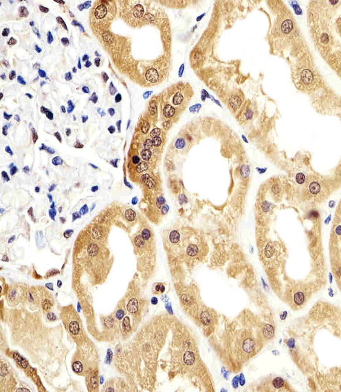
Application
| IHC-P, WB, E |
|---|---|
| Primary Accession | P06733 |
| Reactivity | Human, Mouse |
| Host | Mouse |
| Clonality | Monoclonal |
| Isotype | IgG (k) |
| Clone/Animal Names | 786CT6.6.4 |
| Calculated MW | 47169 Da |
| Gene ID | 2023 |
|---|---|
| Other Names | Alpha-enolase, 2-phospho-D-glycerate hydro-lyase, C-myc promoter-binding protein, Enolase 1, MBP-1, MPB-1, Non-neural enolase, NNE, Phosphopyruvate hydratase, Plasminogen-binding protein, ENO1, ENO1L1, MBPB1, MPB1 |
| Target/Specificity | Purified His-tagged ENO1 protein was used to produced this monoclonal antibody. |
| Dilution | IHC-P~~1:25 WB~~1:1000 E~~Use at an assay dependent concentration. |
| Format | Purified monoclonal antibody supplied in PBS with 0.09% (W/V) sodium azide. This antibody is purified through a protein G column, followed by dialysis against PBS. |
| Storage | Maintain refrigerated at 2-8°C for up to 2 weeks. For long term storage store at -20°C in small aliquots to prevent freeze-thaw cycles. |
| Precautions | ENO1 Antibody is for research use only and not for use in diagnostic or therapeutic procedures. |
| Name | ENO1 |
|---|---|
| Synonyms | ENO1L1, MBPB1, MPB1 |
| Function | Glycolytic enzyme the catalyzes the conversion of 2- phosphoglycerate to phosphoenolpyruvate (PubMed:1369209, PubMed:29775581). In addition to glycolysis, involved in various processes such as growth control, hypoxia tolerance and allergic responses (PubMed:10802057, PubMed:12666133, PubMed:2005901, PubMed:29775581). May also function in the intravascular and pericellular fibrinolytic system due to its ability to serve as a receptor and activator of plasminogen on the cell surface of several cell-types such as leukocytes and neurons (PubMed:12666133). Stimulates immunoglobulin production (PubMed:1369209). |
| Cellular Location | Cytoplasm. Cell membrane. Cytoplasm, myofibril, sarcomere, M line. Note=Can translocate to the plasma membrane in either the homodimeric (alpha/alpha) or heterodimeric (alpha/gamma) form. ENO1 is localized to the M line |
| Tissue Location | The alpha/alpha homodimer is expressed in embryo and in most adult tissues. The alpha/beta heterodimer and the beta/beta homodimer are found in striated muscle, and the alpha/gamma heterodimer and the gamma/gamma homodimer in neurons |

Thousands of laboratories across the world have published research that depended on the performance of antibodies from Abcepta to advance their research. Check out links to articles that cite our products in major peer-reviewed journals, organized by research category.
info@abcepta.com, and receive a free "I Love Antibodies" mug.

Provided below are standard protocols that you may find useful for product applications.
Background
Multifunctional enzyme that, as well as its role in glycolysis, plays a part in various processes such as growth control, hypoxia tolerance and allergic responses. May also function in the intravascular and pericellular fibrinolytic system due to its ability to serve as a receptor and activator of plasminogen on the cell surface of several cell-types such as leukocytes and neurons. Stimulates immunoglobulin production. MBP1 binds to the myc promoter and acts as a transcriptional repressor. May be a tumor suppressor.
References
Giallongo A., et al. Proc. Natl. Acad. Sci. U.S.A. 83:6741-6745(1986).
Giallongo A., et al. Eur. J. Biochem. 190:567-573(1990).
Ray R., et al. Mol. Cell. Biol. 11:2154-2161(1991).
Walter M., et al. J. Autoimmun. 8:931-945(1995).
Kalnine N., et al. Submitted (MAY-2003) to the EMBL/GenBank/DDBJ databases.
If you have used an Abcepta product and would like to share how it has performed, please click on the "Submit Review" button and provide the requested information. Our staff will examine and post your review and contact you if needed.
If you have any additional inquiries please email technical services at tech@abcepta.com.
Ordering Information
Other Products
Shipping Information
