


 Foundational characteristics of cancer include proliferation, angiogenesis, migration, evasion of apoptosis, and cellular immortality. Find key markers for these cellular processes and antibodies to detect them.
 The SUMOplot™ Analysis Program predicts and scores sumoylation sites in your protein. SUMOylation is a post-translational modification involved in various cellular processes, such as nuclear-cytosolic transport, transcriptional regulation, apoptosis, protein stability, response to stress, and progression through the cell cycle.
 The Autophagy Receptor Motif Plotter predicts and scores autophagy receptor binding sites in your protein. Identifying proteins connected to this pathway is critical to understanding the role of autophagy in physiological as well as pathological processes such as development, differentiation, neurodegenerative diseases, stress, infection, and cancer.
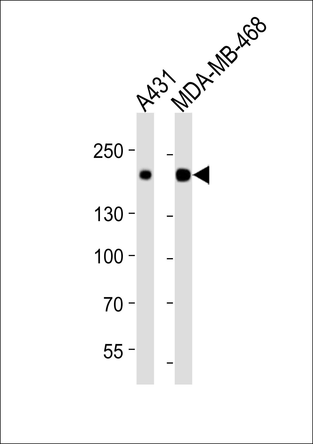
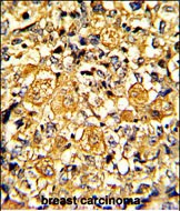
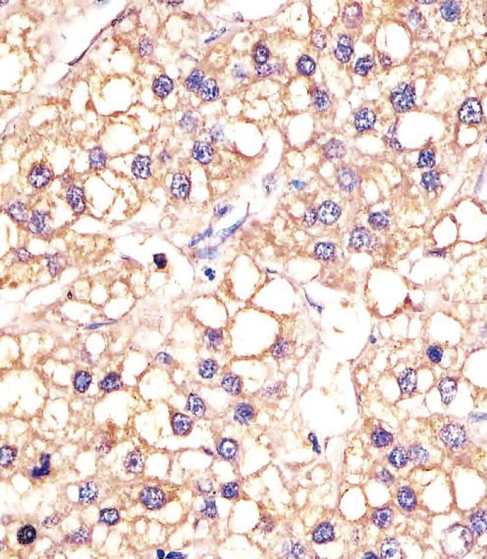
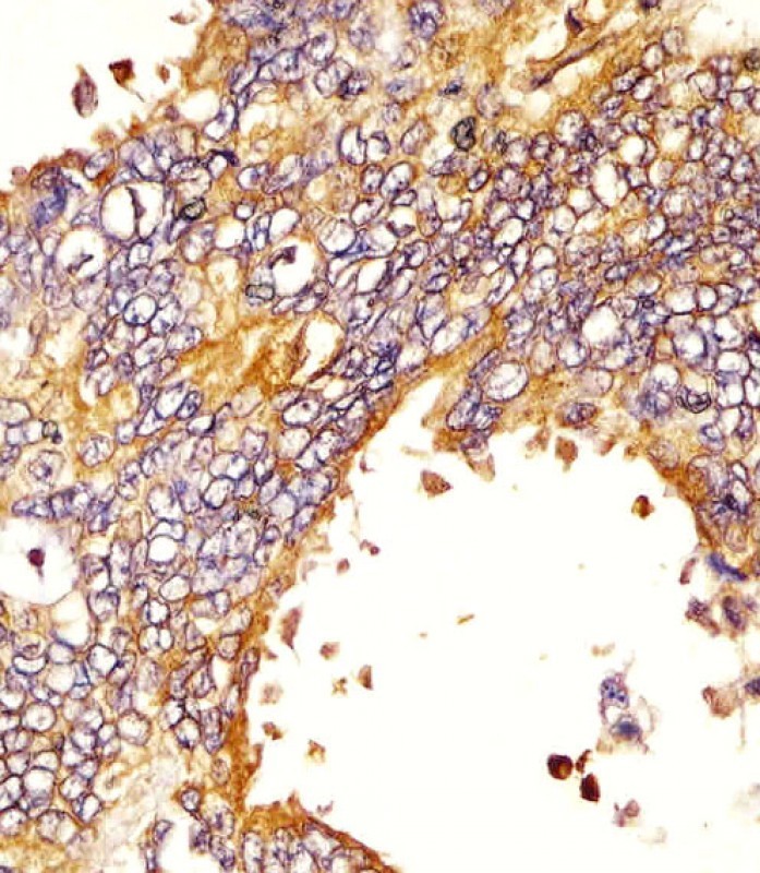
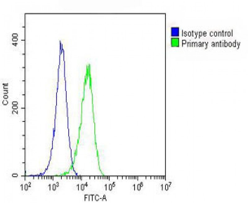
EGFR Antibody
Purified Mouse Monoclonal Antibody (Mab)
- SPECIFICATION
- CITATIONS
- PROTOCOLS
- BACKGROUND

Application
| WB, IHC-P, FC, E |
|---|---|
| Primary Accession | P00533 |
| Reactivity | Human |
| Host | Mouse |
| Clonality | Monoclonal |
| Isotype | IgG1κ |
| Clone/Animal Names | 51CT78.40.5 |
| Calculated MW | 134277 Da |
| Gene ID | 1956 |
|---|---|
| Other Names | Epidermal growth factor receptor, Proto-oncogene c-ErbB-1, Receptor tyrosine-protein kinase erbB-1, EGFR, ERBB, ERBB1, HER1 |
| Target/Specificity | Purified His-tagged EGFR protein(Fragment) was used to produced this monoclonal antibody. |
| Dilution | WB~~1:2000 IHC-P~~1:25 FC~~1:25 E~~Use at an assay dependent concentration. |
| Format | Purified monoclonal antibody supplied in PBS with 0.09% (W/V) sodium azide. This antibody is purified through a protein G column, followed by dialysis against PBS. |
| Storage | Maintain refrigerated at 2-8°C for up to 2 weeks. For long term storage store at -20°C in small aliquots to prevent freeze-thaw cycles. |
| Precautions | EGFR Antibody is for research use only and not for use in diagnostic or therapeutic procedures. |
| Name | EGFR (HGNC:3236) |
|---|---|
| Synonyms | ERBB, ERBB1, HER1 |
| Function | Receptor tyrosine kinase binding ligands of the EGF family and activating several signaling cascades to convert extracellular cues into appropriate cellular responses (PubMed:10805725, PubMed:27153536, PubMed:2790960, PubMed:35538033). Known ligands include EGF, TGFA/TGF- alpha, AREG, epigen/EPGN, BTC/betacellulin, epiregulin/EREG and HBEGF/heparin-binding EGF (PubMed:12297049, PubMed:15611079, PubMed:17909029, PubMed:20837704, PubMed:27153536, PubMed:2790960, PubMed:7679104, PubMed:8144591, PubMed:9419975). Ligand binding triggers receptor homo- and/or heterodimerization and autophosphorylation on key cytoplasmic residues. The phosphorylated receptor recruits adapter proteins like GRB2 which in turn activates complex downstream signaling cascades. Activates at least 4 major downstream signaling cascades including the RAS-RAF-MEK-ERK, PI3 kinase-AKT, PLCgamma-PKC and STATs modules (PubMed:27153536). May also activate the NF-kappa-B signaling cascade (PubMed:11116146). Also directly phosphorylates other proteins like RGS16, activating its GTPase activity and probably coupling the EGF receptor signaling to the G protein-coupled receptor signaling (PubMed:11602604). Also phosphorylates MUC1 and increases its interaction with SRC and CTNNB1/beta-catenin (PubMed:11483589). Positively regulates cell migration via interaction with CCDC88A/GIV which retains EGFR at the cell membrane following ligand stimulation, promoting EGFR signaling which triggers cell migration (PubMed:20462955). Plays a role in enhancing learning and memory performance (By similarity). Plays a role in mammalian pain signaling (long-lasting hypersensitivity) (By similarity). |
| Cellular Location | Cell membrane; Single-pass type I membrane protein. Endoplasmic reticulum membrane; Single-pass type I membrane protein. Golgi apparatus membrane; Single-pass type I membrane protein. Nucleus membrane; Single-pass type I membrane protein Endosome Endosome membrane. Nucleus. Note=In response to EGF, translocated from the cell membrane to the nucleus via Golgi and ER (PubMed:17909029, PubMed:20674546). Endocytosed upon activation by ligand (PubMed:17182860, PubMed:17909029, PubMed:27153536, PubMed:2790960). Colocalized with GPER1 in the nucleus of estrogen agonist-induced cancer-associated fibroblasts (CAF) (PubMed:20551055) |
| Tissue Location | Ubiquitously expressed. Isoform 2 is also expressed in ovarian cancers. |

Thousands of laboratories across the world have published research that depended on the performance of antibodies from Abcepta to advance their research. Check out links to articles that cite our products in major peer-reviewed journals, organized by research category.
info@abcepta.com, and receive a free "I Love Antibodies" mug.

Provided below are standard protocols that you may find useful for product applications.
Background
The protein encoded by this gene is a transmembrane glycoprotein that is a member of the protein kinase superfamily. This protein is a receptor for members of the epidermal growth factor family. EGFR is a cell surface protein that binds to epidermal growth factor. Binding of the protein to a ligand induces receptor dimerization and tyrosine autophosphorylation and leads to cell proliferation. Mutations in this gene are associated with lung cancer. Multiple alternatively spliced transcript variants that encode different protein isoforms have been found for this gene.
References
Complex Mutations in the Epidermal Growth Factor Receptor Gene in Non-small Cell Lung Cancer. Hata A, et al. J Thorac Oncol, 2010 Aug 30. PMID 20808254. EGFR signaling is differentially activated in patient-derived glioblastoma stem cells. Howard BM, et al. J Exp Ther Oncol, 2010. PMID 20734923. [EGFR Mutations Detection in Non-small Cell Lung Cancer Tissues by Real-time PCR and DNA Sequencing.] Li Y, et al. Zhongguo Fei Ai Za Zhi, 2009 Dec 20. PMID 20723379. [Detection and Its Clinical Significance of EGFR Gene Mutation and Gene Amplification in 187 Patients with Non-small Cell Lung Cancer.] Liu H, et al. Zhongguo Fei Ai Za Zhi, 2009 Dec 20. PMID 20723374. Effect of gefitinib on the survival of patients with recurrence of lung adenocarcinoma after surgery: A retrospective case-matching cohort study. Katayama T, et al. Surg Oncol, 2010 Aug 10. PMID 20705455.
If you have used an Abcepta product and would like to share how it has performed, please click on the "Submit Review" button and provide the requested information. Our staff will examine and post your review and contact you if needed.
If you have any additional inquiries please email technical services at tech@abcepta.com.
Ordering Information
Other Products
Shipping Information
