


 Foundational characteristics of cancer include proliferation, angiogenesis, migration, evasion of apoptosis, and cellular immortality. Find key markers for these cellular processes and antibodies to detect them.
 The SUMOplot™ Analysis Program predicts and scores sumoylation sites in your protein. SUMOylation is a post-translational modification involved in various cellular processes, such as nuclear-cytosolic transport, transcriptional regulation, apoptosis, protein stability, response to stress, and progression through the cell cycle.
 The Autophagy Receptor Motif Plotter predicts and scores autophagy receptor binding sites in your protein. Identifying proteins connected to this pathway is critical to understanding the role of autophagy in physiological as well as pathological processes such as development, differentiation, neurodegenerative diseases, stress, infection, and cancer.
> home > Products > Primary Antibodies > Antibody Collections > Mitochondrion > HK2 (Hexokinase II) Antibody
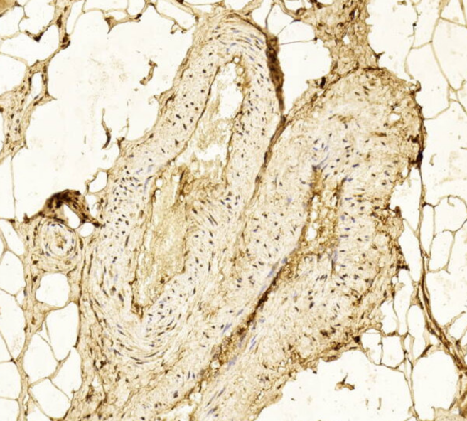
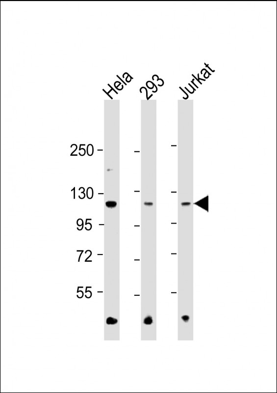
HK2 (Hexokinase II) Antibody
Purified Mouse Monoclonal Antibody (Mab)
- SPECIFICATION
- CITATIONS: 1
- PROTOCOLS
- BACKGROUND

Application
| WB, IHC-P, E |
|---|---|
| Primary Accession | P52789 |
| Reactivity | Human |
| Host | Mouse |
| Clonality | monoclonal |
| Isotype | IgG1,k |
| Clone/Animal Names | 1798CT748.15.25 |
| Calculated MW | 102380 Da |
| Gene ID | 3099 |
|---|---|
| Other Names | Hexokinase-2, 2.7.1.1, Hexokinase type II, HK II, Muscle form hexokinase, HK2 |
| Target/Specificity | This HK2 (Hexokinase II) antibody is generated from a mouse immunized with a recombinant protein between 1-170 amino acids from human HK2 (Hexokinase II). |
| Dilution | WB~~1:500-1:1000 IHC-P~~N/A E~~Use at an assay dependent concentration. |
| Format | Purified monoclonal antibody supplied in PBS with 0.09% (W/V) sodium azide. This antibody is purified through a protein G column, followed by dialysis against PBS. |
| Storage | Maintain refrigerated at 2-8°C for up to 2 weeks. For long term storage store at -20°C in small aliquots to prevent freeze-thaw cycles. |
| Precautions | HK2 (Hexokinase II) Antibody is for research use only and not for use in diagnostic or therapeutic procedures. |
| Name | HK2 (HGNC:4923) |
|---|---|
| Function | Catalyzes the phosphorylation of hexose, such as D-glucose and D-fructose, to hexose 6-phosphate (D-glucose 6-phosphate and D- fructose 6-phosphate, respectively) (PubMed:23185017, PubMed:26985301, PubMed:29298880). Mediates the initial step of glycolysis by catalyzing phosphorylation of D-glucose to D-glucose 6-phosphate (PubMed:29298880). Plays a key role in maintaining the integrity of the outer mitochondrial membrane by preventing the release of apoptogenic molecules from the intermembrane space and subsequent apoptosis (PubMed:18350175). |
| Cellular Location | Mitochondrion outer membrane; Peripheral membrane protein. Cytoplasm, cytosol Note=The mitochondrial-binding peptide (MBP) region promotes association with the mitochondrial outer membrane (PubMed:29298880) The interaction with the mitochondrial outer membrane via the mitochondrial-binding peptide (MBP) region promotes higher stability of the protein (PubMed:29298880). Release from the mitochondrial outer membrane into the cytosol induces permeability transition pore (PTP) opening and apoptosis (PubMed:18350175). |
| Tissue Location | Predominant hexokinase isozyme expressed in insulin-responsive tissues such as skeletal muscle |
Research Areas
Citations ( 0 )

Application Protocols
Provided below are standard protocols that you may find useful for product applications.
References
Deeb S.S.,et al.Biochem. Biophys. Res. Commun. 197:68-74(1993).
Lehto M.,et al.Diabetologia 38:1466-1474(1995).
Malkki M.,et al.Submitted (MAY-1999) to the EMBL/GenBank/DDBJ databases.
Mural R.J.,et al.Submitted (SEP-2005) to the EMBL/GenBank/DDBJ databases.
Shinohara Y.,et al.Cancer Lett. 82:27-32(1994).
Abcepta welcomes feedback from its customers.
If you have used an Abcepta product and would like to share how it has performed, please click on the "Submit Review" button and provide the requested information. Our staff will examine and post your review and contact you if needed.
If you have any additional inquiries please email technical services at tech@abcepta.com.
$ 150.00
$ 385.00
Cat# AM8606b
Ordering Information
United States
AlbaniaAustraliaAustriaBelgiumBosnia & HerzegovinaBrazilBulgariaCanadaCentral AmericaChinaCroatiaCyprusCzech RepublicDenmarkEstoniaFinlandFranceGermanyGreeceHong KongHungaryIcelandIndiaIndonesiaIrelandIsraelItalyJapanLatviaLithuaniaLuxembourgMacedoniaMalaysiaMaltaMexicoNetherlandsNew ZealandNorwayPakistanPolandPortugalRomaniaSerbiaSingaporeSlovakiaSloveniaSouth AfricaSouth KoreaSpainSwedenSwitzerlandTaiwanTurkeyUnited KingdomUnited StatesVietnamWorldwideOthers
USA Headquarters
(888) 735-7227 / (858) 622-0099 or (858) 875-1900
Other Products
Shipping Information
Domestic orders (in stock items)
Shipped out the same day. Orders placed after 1 PM (PST) will ship out the next business day.
International orders
Contact your local distributors
