


 Foundational characteristics of cancer include proliferation, angiogenesis, migration, evasion of apoptosis, and cellular immortality. Find key markers for these cellular processes and antibodies to detect them.
 The SUMOplot™ Analysis Program predicts and scores sumoylation sites in your protein. SUMOylation is a post-translational modification involved in various cellular processes, such as nuclear-cytosolic transport, transcriptional regulation, apoptosis, protein stability, response to stress, and progression through the cell cycle.
 The Autophagy Receptor Motif Plotter predicts and scores autophagy receptor binding sites in your protein. Identifying proteins connected to this pathway is critical to understanding the role of autophagy in physiological as well as pathological processes such as development, differentiation, neurodegenerative diseases, stress, infection, and cancer.
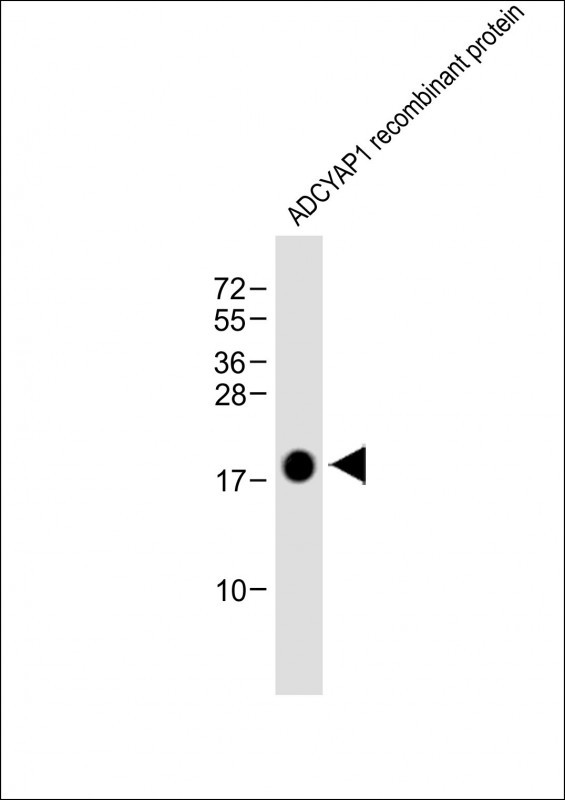
ADCYAP1 Antibody
Purified Mouse Monoclonal Antibody (Mab)
- SPECIFICATION
- CITATIONS
- PROTOCOLS
- BACKGROUND

Application
| WB, E |
|---|---|
| Primary Accession | P18509 |
| Reactivity | Human |
| Predicted | Human |
| Host | Mouse |
| Clonality | monoclonal |
| Isotype | IgG1,κ |
| Clone/Animal Names | 1996CT329.45.62 |
| Calculated MW | 18835 Da |
| Gene ID | 116 |
|---|---|
| Other Names | Pituitary adenylate cyclase-activating polypeptide, PACAP, PACAP-related peptide, PRP-48, Pituitary adenylate cyclase-activating polypeptide 27, PACAP-27, PACAP27, Pituitary adenylate cyclase-activating polypeptide 38, PACAP-38, PACAP38, ADCYAP1 |
| Target/Specificity | This ADCYAP1 antibody is generated from a mouse immunized with a recombinant protein from the human region of human ADCYAP1. |
| Dilution | WB~~1:8000 E~~Use at an assay dependent concentration. |
| Format | Purified monoclonal antibody supplied in PBS with 0.09% (W/V) sodium azide. This antibody is purified through a protein G column, followed by dialysis against PBS. |
| Storage | Maintain refrigerated at 2-8°C for up to 2 weeks. For long term storage store at -20°C in small aliquots to prevent freeze-thaw cycles. |
| Precautions | ADCYAP1 Antibody is for research use only and not for use in diagnostic or therapeutic procedures. |
| Name | ADCYAP1 (HGNC:241) |
|---|---|
| Function | PACAP is a neuropeptide involved in diverse array of physiological processes through activating the PACAP subfamily of class B1 G protein-coupled receptors: VIP receptor 1 (VIPR1), VIP receptor 2 (VIPR2), and PACAP type I receptor (ADCYAP1R1) (PubMed:11175907, PubMed:23800469, PubMed:32047270, PubMed:36385145). Exerts neuroprotective and general cytoprotective effects due to anti- apoptotic, anti-inflammatory, and antioxidant actions (PubMed:23800469). Promotes neuron projection development through the RAPGEF2/Rap1/B-Raf/ERK pathway (PubMed:23800469). In chromaffin cells, induces long-lasting increase of intracellular calcium concentrations and neuroendocrine secretion (By similarity). Involved in the control of glucose homeostasis, induces insulin secretion by pancreatic beta cells (By similarity). PACAP exists in two bioactive forms from proteolysis of the same precursor protein, PACAP27 and PACAP38, which differ by eleven amino acid residues in the C-terminus (PubMed:32047270). |
| Cellular Location | Secreted. |

Thousands of laboratories across the world have published research that depended on the performance of antibodies from Abcepta to advance their research. Check out links to articles that cite our products in major peer-reviewed journals, organized by research category.
info@abcepta.com, and receive a free "I Love Antibodies" mug.

Provided below are standard protocols that you may find useful for product applications.
Background
Binding to its receptor activates G proteins and stimulates adenylate cyclase in pituitary cells. Promotes neuron projection development through the RAPGEF2/Rap1/B-Raf/ERK pathway.
References
Ohkubo S.,et al.DNA Cell Biol. 11:21-30(1992).
Hosoya M.,et al.Biochim. Biophys. Acta 1129:199-206(1992).
Ota T.,et al.Nat. Genet. 36:40-45(2004).
Kimura C.,et al.Biochem. Biophys. Res. Commun. 166:81-89(1990).
Emery A.C.,et al.Sci. Signal. 6:RA51-RA51(2013).
If you have used an Abcepta product and would like to share how it has performed, please click on the "Submit Review" button and provide the requested information. Our staff will examine and post your review and contact you if needed.
If you have any additional inquiries please email technical services at tech@abcepta.com.
Ordering Information
Other Products
Shipping Information
