


 Foundational characteristics of cancer include proliferation, angiogenesis, migration, evasion of apoptosis, and cellular immortality. Find key markers for these cellular processes and antibodies to detect them.
 The SUMOplot™ Analysis Program predicts and scores sumoylation sites in your protein. SUMOylation is a post-translational modification involved in various cellular processes, such as nuclear-cytosolic transport, transcriptional regulation, apoptosis, protein stability, response to stress, and progression through the cell cycle.
 The Autophagy Receptor Motif Plotter predicts and scores autophagy receptor binding sites in your protein. Identifying proteins connected to this pathway is critical to understanding the role of autophagy in physiological as well as pathological processes such as development, differentiation, neurodegenerative diseases, stress, infection, and cancer.
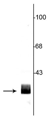
Anti-14-3-3 Protein Antibody
Our Anti-14-3-3 Protein rabbit polyclonal primary antibody from PhosphoSolutions is produced in-hous
- SPECIFICATION
- CITATIONS
- PROTOCOLS
- BACKGROUND

| Primary Accession | P35213 |
|---|---|
| Reactivity | Bovine |
| Host | Rabbit |
| Clonality | Polyclonal |
| Isotype | IgG |
| Calculated MW | 28054 Da |
| Gene ID | 56011 |
|---|---|
| Other Names | 14 3 3 antibody, 14 3 3 protein beta antibody, 14 3 3 protein beta/alpha antibody, 14 3 3 protein zeta antibody , 14 3 3 zeta antibody, 14-3-3 protein beta/alpha antibody, 14-3-3 protein/cytosolic phospholipase A2 antibody, 1433B_HUMAN antibody, GW128 antibody, HS1 antibody, KCIP 1 antibody, KCIP-1 antibody, MGC111427 antibody, MGC126532 antibody, MGC138156 antibody, N-terminally processed antibody, Protein 1054 antibody, Protein kinase C inhibitor protein 1 antibody, Tyrosine 3-monooxygenase/tryptophan 5-monooxygenase activation protein delta polypeptide antibody, Tyrosine 3/tryptophan 5 -monooxygenase activation protein zeta polypeptide antibody, YWHAB antibody, YWHAD antibody, YWHAZ antibody |
| Target/Specificity | 14-3-3 proteins are a family of highly conserved proteins that appear to have multiple roles in cell signaling (Bridges and Moorhead, 2005). The proteins are abundantly expressed in the brain and have been detected in the cerebrospinal fluid of patients with different neurological disorders (Berg et al., 2003). 14-3-3 proteins bind protein ligands that are typically phosphorylated on serine or threonine residues and regulate the functions of these binding partners by a number of different mechanisms (Silhan et al., 2004; Dougherty and Morrison, 2004). The 14-3-3 proteins affect a diverse array of cellular processes including the cell cycle and transcription, signal transduction and intracellular trafficking. |
| Format | Antigen Affinity Purified from Pooled Serum |
| Storage | Maintain refrigerated at 2-8°C for up to 6 months. For long term storage store at -20°C in small aliquots to prevent freeze-thaw cycles. |
| Precautions | Anti-14-3-3 Protein Antibody is for research use only and not for use in diagnostic or therapeutic procedures. |
| Shipping | Blue Ice |

Thousands of laboratories across the world have published research that depended on the performance of antibodies from Abcepta to advance their research. Check out links to articles that cite our products in major peer-reviewed journals, organized by research category.
info@abcepta.com, and receive a free "I Love Antibodies" mug.

Provided below are standard protocols that you may find useful for product applications.
Background
14-3-3 proteins are a family of highly conserved proteins that appear to have multiple roles in cell signaling (Bridges and Moorhead, 2005). The proteins are abundantly expressed in the brain and have been detected in the cerebrospinal fluid of patients with different neurological disorders (Berg et al., 2003). 14-3-3 proteins bind protein ligands that are typically phosphorylated on serine or threonine residues and regulate the functions of these binding partners by a number of different mechanisms (Silhan et al., 2004; Dougherty and Morrison, 2004). The 14-3-3 proteins affect a diverse array of cellular processes including the cell cycle and transcription, signal transduction and intracellular trafficking.
If you have used an Abcepta product and would like to share how it has performed, please click on the "Submit Review" button and provide the requested information. Our staff will examine and post your review and contact you if needed.
If you have any additional inquiries please email technical services at tech@abcepta.com.
Ordering Information
Other Products
Shipping Information
