


 Foundational characteristics of cancer include proliferation, angiogenesis, migration, evasion of apoptosis, and cellular immortality. Find key markers for these cellular processes and antibodies to detect them.
 The SUMOplot™ Analysis Program predicts and scores sumoylation sites in your protein. SUMOylation is a post-translational modification involved in various cellular processes, such as nuclear-cytosolic transport, transcriptional regulation, apoptosis, protein stability, response to stress, and progression through the cell cycle.
 The Autophagy Receptor Motif Plotter predicts and scores autophagy receptor binding sites in your protein. Identifying proteins connected to this pathway is critical to understanding the role of autophagy in physiological as well as pathological processes such as development, differentiation, neurodegenerative diseases, stress, infection, and cancer.
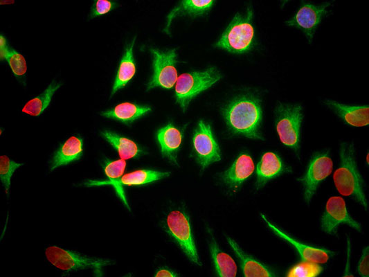
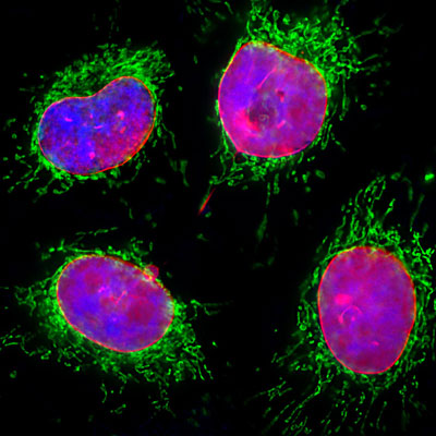
Anti-Lamin A/C Antibody
Our Anti-Lamin A/C primary antibody from PhosphoSolutions is mouse monoclonal. It detects human Lami
- SPECIFICATION
- CITATIONS
- PROTOCOLS
- BACKGROUND

Application
| WB, IHC |
|---|---|
| Primary Accession | P02545 |
| Host | Mouse |
| Clonality | Monoclonal |
| Isotype | IgG1 |
| Clone Names | 4C4 |
| Calculated MW | 74139 Da |
| Gene ID | 4000 |
|---|---|
| Other Names | 70 kDa lamin antibody, Cardiomyopathy dilated 1A (autosomal dominant) antibody, CDCD1 antibody, CDDC antibody, CMD1A antibody, CMT2B1 antibody, EMD2 antibody, FPL antibody, FPLD antibody, FPLD2 antibody, HGPS antibody, IDC antibody, Lamin A antibody, Lamin A/C antibody, Lamin A/C like 1 antibody, Lamin antibody, Lamin C antibody, Lamin-A/C antibody, LDP1 antibody, LFP antibody, LGMD1B antibody, Limb girdle muscular dystrophy 1B (autosomal dominant) antibody, LMN 1 antibody, LMN A antibody, LMN C antibody, LMN1 antibody, LMNA antibody, LMNA_HUMAN antibody, LMNC antibody, LMNL1 antibody, Prelamin A/C antibody, PRO1 antibody, Renal carcinoma antigen NY REN 32 antibody, Renal carcinoma antigen NY-REN-32 antibody, Renal carcinoma antigen NYREN32 antibody |
| Target/Specificity | Lamins A and C are nuclear structural proteins that are part of the intermediate filament family and coded for by the same gene (LMNA). Lamins A and C are nearly identical except for their carboxy termini (McKeon et al., 1986). Mutations in the gene encoding lamins A/C have been shown to cause a variety of diseases including autosomal dominant Emery-Dreifuss muscular dystrophy (Bonne et al., 1995), autosomal dominant limbgirdle muscular dystrophy (Muchir et al., 2000) and Charcot-Marie-Tooth disorder type 2 (De Sandre-Giavonnoli et al., 2002). |
| Dilution | WB~~1:1000 IHC~~1:100~500 |
| Format | Protein G Purified |
| Storage | Maintain refrigerated at 2-8°C for up to 6 months. For long term storage store at -20°C in small aliquots to prevent freeze-thaw cycles. |
| Precautions | Anti-Lamin A/C Antibody is for research use only and not for use in diagnostic or therapeutic procedures. |
| Shipping | Blue Ice |

Thousands of laboratories across the world have published research that depended on the performance of antibodies from Abcepta to advance their research. Check out links to articles that cite our products in major peer-reviewed journals, organized by research category.
info@abcepta.com, and receive a free "I Love Antibodies" mug.

Provided below are standard protocols that you may find useful for product applications.
Background
Lamins A and C are nuclear structural proteins that are part of the intermediate filament family and coded for by the same gene (LMNA). Lamins A and C are nearly identical except for their carboxy termini (McKeon et al., 1986). Mutations in the gene encoding lamins A/C have been shown to cause a variety of diseases including autosomal dominant Emery-Dreifuss muscular dystrophy (Bonne et al., 1995), autosomal dominant limbgirdle muscular dystrophy (Muchir et al., 2000) and Charcot-Marie-Tooth disorder type 2 (De Sandre-Giavonnoli et al., 2002).
If you have used an Abcepta product and would like to share how it has performed, please click on the "Submit Review" button and provide the requested information. Our staff will examine and post your review and contact you if needed.
If you have any additional inquiries please email technical services at tech@abcepta.com.
Ordering Information
Other Products
Shipping Information
