


 Foundational characteristics of cancer include proliferation, angiogenesis, migration, evasion of apoptosis, and cellular immortality. Find key markers for these cellular processes and antibodies to detect them.
 The SUMOplot™ Analysis Program predicts and scores sumoylation sites in your protein. SUMOylation is a post-translational modification involved in various cellular processes, such as nuclear-cytosolic transport, transcriptional regulation, apoptosis, protein stability, response to stress, and progression through the cell cycle.
 The Autophagy Receptor Motif Plotter predicts and scores autophagy receptor binding sites in your protein. Identifying proteins connected to this pathway is critical to understanding the role of autophagy in physiological as well as pathological processes such as development, differentiation, neurodegenerative diseases, stress, infection, and cancer.
Anti-Peripherin Antibody
Our Anti-Peripherin primary antibody from PhosphoSolutions is mouse monoclonal. It detects mouse and
- SPECIFICATION
- CITATIONS
- PROTOCOLS
- BACKGROUND

Application
| WB, IHC |
|---|---|
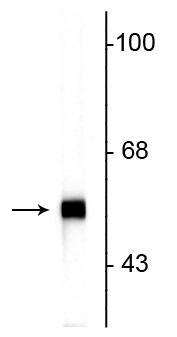
| Primary Accession | P21807 |
| Host | Mouse |
| Clonality | Monoclonal |
| Isotype | IgG1 |
| Clone Names | 7C5 |
| Calculated MW | 53550 Da |
| Other Names | NEF 4 antibody, NEF4 antibody, Neurofilament 4 (57kD) antibody, Neurofilament 4 antibody, Perf antibody, PERI_HUMAN antibody, Peripherin antibody, PRPH 1 antibody, prph antibody, PRPH1 antibody |
|---|---|
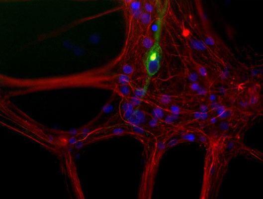
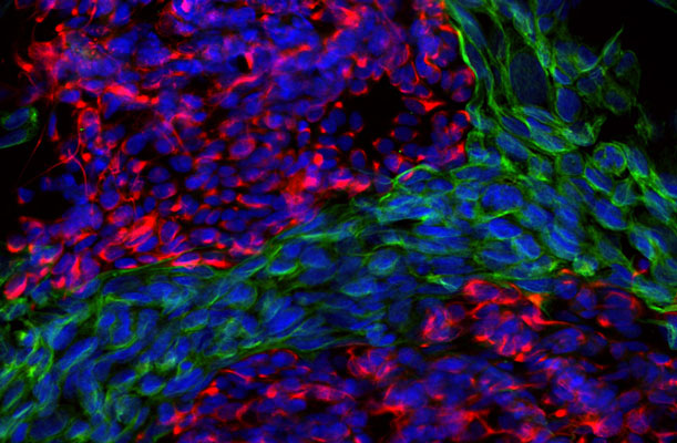
| Target/Specificity | Peripherin is a ~57 kDa intermediate filament subunit found initially in sensory neurons of the peripheral nervous systems, which gives the protein its name. Subsequently, peripherin was found in some sensory and other neurons of the central nervous system and also in PC12 cells. Peripherin is also expressed in certain neuroendocrine tumors and in the insulin producing cells of the pancreas. Peripherin belongs to the Class III family of intermediate filament subunits which also includes vimentin, glial fibrillary acidic protein (GFAP) and desmin. In contrast to the neurofilaments, peripherin is strongly up-regulated after nerve injury (1). Antibodies to peripherin can be used in identifying, classifying, and studying neurons throughout the nervous system. Peripherin is also a good diagnostic marker for ballooned axons seen in Lou Gehrig's disease (Amyotrophic lateral sclerosis) and some neuronally derived tumors (2). Autoantibodies to peripherin are frequently seen in the sera of patients with diabetes (3). Peripherin is not related to peripherin/RDS, a protein of the photoreceptor outer membrane mutations of which are causative of certain forms of slow retinal degeneration. |
| Dilution | WB~~1:1000 IHC~~1:100~500 |
| Format | Concentrated tissue culture supernatant |
| Storage | Maintain refrigerated at 2-8°C for up to 6 months. For long term storage store at -20°C in small aliquots to prevent freeze-thaw cycles. |
| Precautions | Anti-Peripherin Antibody is for research use only and not for use in diagnostic or therapeutic procedures. |
| Shipping | Blue Ice |

Thousands of laboratories across the world have published research that depended on the performance of antibodies from Abcepta to advance their research. Check out links to articles that cite our products in major peer-reviewed journals, organized by research category.
info@abcepta.com, and receive a free "I Love Antibodies" mug.

Provided below are standard protocols that you may find useful for product applications.
Background
Peripherin is a ~57 kDa intermediate filament subunit found initially in sensory neurons of the peripheral nervous systems, which gives the protein its name. Subsequently, peripherin was found in some sensory and other neurons of the central nervous system and also in PC12 cells. Peripherin is also expressed in certain neuroendocrine tumors and in the insulin producing cells of the pancreas. Peripherin belongs to the Class III family of intermediate filament subunits which also includes vimentin, glial fibrillary acidic protein (GFAP) and desmin. In contrast to the neurofilaments, peripherin is strongly up-regulated after nerve injury (1). Antibodies to peripherin can be used in identifying, classifying, and studying neurons throughout the nervous system. Peripherin is also a good diagnostic marker for ballooned axons seen in Lou Gehrig's disease (Amyotrophic lateral sclerosis) and some neuronally derived tumors (2). Autoantibodies to peripherin are frequently seen in the sera of patients with diabetes (3). Peripherin is not related to peripherin/RDS, a protein of the photoreceptor outer membrane mutations of which are causative of certain forms of slow retinal degeneration.
If you have used an Abcepta product and would like to share how it has performed, please click on the "Submit Review" button and provide the requested information. Our staff will examine and post your review and contact you if needed.
If you have any additional inquiries please email technical services at tech@abcepta.com.
Ordering Information
Other Products
Shipping Information
