


 Foundational characteristics of cancer include proliferation, angiogenesis, migration, evasion of apoptosis, and cellular immortality. Find key markers for these cellular processes and antibodies to detect them.
 The SUMOplot™ Analysis Program predicts and scores sumoylation sites in your protein. SUMOylation is a post-translational modification involved in various cellular processes, such as nuclear-cytosolic transport, transcriptional regulation, apoptosis, protein stability, response to stress, and progression through the cell cycle.
 The Autophagy Receptor Motif Plotter predicts and scores autophagy receptor binding sites in your protein. Identifying proteins connected to this pathway is critical to understanding the role of autophagy in physiological as well as pathological processes such as development, differentiation, neurodegenerative diseases, stress, infection, and cancer.
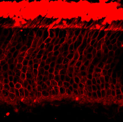
Anti-Rhodopsin Antibody
Our Anti-Rhodopsin primary antibody from PhosphoSolutions is mouse monoclonal. It detects amphibians
- SPECIFICATION
- CITATIONS
- PROTOCOLS
- BACKGROUND

Application
| WB |
|---|---|
| Primary Accession | P02699 |
| Host | Mouse |
| Clonality | Monoclonal |
| Isotype | IgG1 |
| Clone Names | 1D4 |
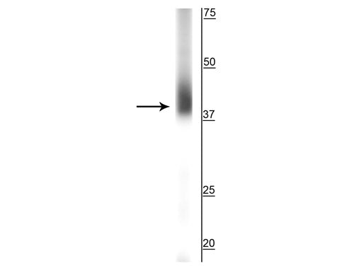
| Calculated MW | 39008 Da |
| Gene ID | 509933 |
|---|---|
| Other Names | CSNBAD1 antibody, MGC138309 antibody, MGC138311 antibody, OPN 2 antibody, OPN2 antibody, opsd antibody, OPSD_HUMAN antibody, opsin 2 antibody, Opsin 2 rod pigment antibody, Opsin-2 antibody, Opsin2 antibody, Retinitis Pigmentosa 4 antibody, Retinitis pigmentosa 4 autosomal dominant antibody, RHO antibody, Rhodopsin antibody, RP 4 antibody, RP4 antibody |
| Target/Specificity | Rhodopsin is a photoreceptor protein found in retinal rods. It is a complex formed by the binding of retinaldehyde, the oxidized form of retinol, to the protein opsin and undergoes a series of complex reactions in response to visible light resulting in the transmission of nerve impulses to the brain. Mutation of the rhodopsin gene is a major contributor to various retinopathies such as retinitis pigmentosa. The disease-causing protein generally aggregates with ubiquitin in inclusion bodies, disrupts the intermediate filament network and impairs the ability of the cell to degrade non-functioning proteins which leads to photoreceptor apoptosis (Berson et al., 1991). Other mutations on rhodopsin lead to X-linked congenital stationary night blindness, mainly due to constitutive activation, when the mutations occur around the chromophore binding pocket of rhodopsin (Dryja et al.,1993). Several other pathological states relating to rhodopsin have been discovered including poor post-Golgi trafficking, dysregulative activation, rod outer segment instability and arrestin binding. |
| Dilution | WB~~1:1000 |
| Format | Protein G Purified |
| Storage | Maintain refrigerated at 2-8°C for up to 6 months. For long term storage store at -20°C in small aliquots to prevent freeze-thaw cycles. |
| Precautions | Anti-Rhodopsin Antibody is for research use only and not for use in diagnostic or therapeutic procedures. |
| Shipping | Blue Ice |

Thousands of laboratories across the world have published research that depended on the performance of antibodies from Abcepta to advance their research. Check out links to articles that cite our products in major peer-reviewed journals, organized by research category.
info@abcepta.com, and receive a free "I Love Antibodies" mug.

Provided below are standard protocols that you may find useful for product applications.
Background
Rhodopsin is a photoreceptor protein found in retinal rods. It is a complex formed by the binding of retinaldehyde, the oxidized form of retinol, to the protein opsin and undergoes a series of complex reactions in response to visible light resulting in the transmission of nerve impulses to the brain. Mutation of the rhodopsin gene is a major contributor to various retinopathies such as retinitis pigmentosa. The disease-causing protein generally aggregates with ubiquitin in inclusion bodies, disrupts the intermediate filament network and impairs the ability of the cell to degrade non-functioning proteins which leads to photoreceptor apoptosis (Berson et al., 1991). Other mutations on rhodopsin lead to X-linked congenital stationary night blindness, mainly due to constitutive activation, when the mutations occur around the chromophore binding pocket of rhodopsin (Dryja et al.,1993). Several other pathological states relating to rhodopsin have been discovered including poor post-Golgi trafficking, dysregulative activation, rod outer segment instability and arrestin binding.
If you have used an Abcepta product and would like to share how it has performed, please click on the "Submit Review" button and provide the requested information. Our staff will examine and post your review and contact you if needed.
If you have any additional inquiries please email technical services at tech@abcepta.com.
Ordering Information
Other Products
Shipping Information
