


 Foundational characteristics of cancer include proliferation, angiogenesis, migration, evasion of apoptosis, and cellular immortality. Find key markers for these cellular processes and antibodies to detect them.
 The SUMOplot™ Analysis Program predicts and scores sumoylation sites in your protein. SUMOylation is a post-translational modification involved in various cellular processes, such as nuclear-cytosolic transport, transcriptional regulation, apoptosis, protein stability, response to stress, and progression through the cell cycle.
 The Autophagy Receptor Motif Plotter predicts and scores autophagy receptor binding sites in your protein. Identifying proteins connected to this pathway is critical to understanding the role of autophagy in physiological as well as pathological processes such as development, differentiation, neurodegenerative diseases, stress, infection, and cancer.
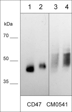
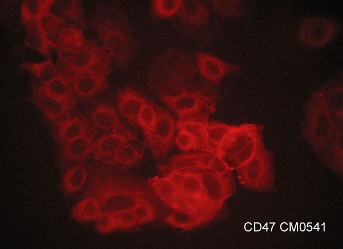
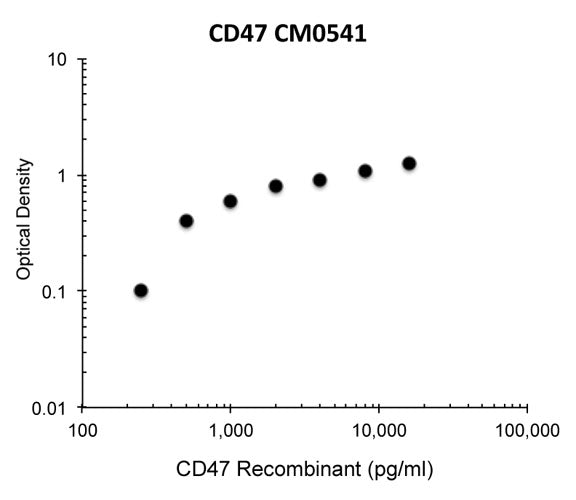
Anti-CD47 (Extracellular region) Antibody
- SPECIFICATION
- CITATIONS
- PROTOCOLS
- BACKGROUND

Application
| WB |
|---|---|
| Primary Accession | Q08722 |
| Host | Mouse |
| Clonality | Mouse Monoclonal |
| Isotype | IgG1 |
| Clone Names | M054 |
| Calculated MW | 35214 Da |
| Gene ID | 961 |
|---|---|
| Other Names | Antigenic surface determinant protein OA3, Integrin-associated protein, IAP, Leukocyte surface antigen CD47, MER6 |
| Target/Specificity | CD47 is a five-pass transmembrane protein expressed on all normal cells, as well as in cancer cells. CD47 is used by macrophages to distinguish between "self" and "non-self" cells. SIRPα expressed on myeloid cells including macrophages, and neuronal cells in the central nervous system, can bind CD47. SIRPα cytoplasmic tail can inhibit macrophage phagocytosis towards CD47-expressing cells. Thus, the CD47/SIRPα pahtway serves as an innate immune checkpoint. Additionally, CD47 was reported to modulate lymphocyte cell activation and proliferation. CD47 is over-expressed in many types of cancer, and the expression level of CD47 on cancer cells is negatively associated with cancer survival. Monoclonal antibody therapies that can block CD47-SIRPα interaction are being actively pursued for clinical applications. In addition to SIRPα, CD47 interacts with thrombospondin-1, VEGFR2, FAS, and certain integrins in different contexts, and influences their downstream signaling. |
| Dilution | WB~~1:1000 |
| Format | Protein G Purified |
| Storage | Maintain refrigerated at 2-8°C for up to 6 months. For long term storage store at -20°C in small aliquots to prevent freeze-thaw cycles. |
| Precautions | Anti-CD47 (Extracellular region) Antibody is for research use only and not for use in diagnostic or therapeutic procedures. |
| Shipping | Blue Ice |

Thousands of laboratories across the world have published research that depended on the performance of antibodies from Abcepta to advance their research. Check out links to articles that cite our products in major peer-reviewed journals, organized by research category.
info@abcepta.com, and receive a free "I Love Antibodies" mug.

Provided below are standard protocols that you may find useful for product applications.
Background
CD47 is a five-pass transmembrane protein expressed on all normal cells, as well as in cancer cells. CD47 is used by macrophages to distinguish between "self" and "non-self" cells. SIRPα expressed on myeloid cells including macrophages, and neuronal cells in the central nervous system, can bind CD47. SIRPα cytoplasmic tail can inhibit macrophage phagocytosis towards CD47-expressing cells. Thus, the CD47/SIRPα pahtway serves as an innate immune checkpoint. Additionally, CD47 was reported to modulate lymphocyte cell activation and proliferation. CD47 is over-expressed in many types of cancer, and the expression level of CD47 on cancer cells is negatively associated with cancer survival. Monoclonal antibody therapies that can block CD47-SIRPα interaction are being actively pursued for clinical applications. In addition to SIRPα, CD47 interacts with thrombospondin-1, VEGFR2, FAS, and certain integrins in different contexts, and influences their downstream signaling.
If you have used an Abcepta product and would like to share how it has performed, please click on the "Submit Review" button and provide the requested information. Our staff will examine and post your review and contact you if needed.
If you have any additional inquiries please email technical services at tech@abcepta.com.
Ordering Information
Other Products
Shipping Information
