


 Foundational characteristics of cancer include proliferation, angiogenesis, migration, evasion of apoptosis, and cellular immortality. Find key markers for these cellular processes and antibodies to detect them.
 The SUMOplot™ Analysis Program predicts and scores sumoylation sites in your protein. SUMOylation is a post-translational modification involved in various cellular processes, such as nuclear-cytosolic transport, transcriptional regulation, apoptosis, protein stability, response to stress, and progression through the cell cycle.
 The Autophagy Receptor Motif Plotter predicts and scores autophagy receptor binding sites in your protein. Identifying proteins connected to this pathway is critical to understanding the role of autophagy in physiological as well as pathological processes such as development, differentiation, neurodegenerative diseases, stress, infection, and cancer.
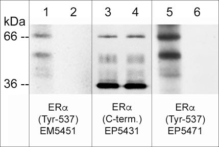
Anti-Estrogen Receptor α (Tyr-537), Phosphospecific Antibody
- SPECIFICATION
- CITATIONS
- PROTOCOLS
- BACKGROUND

| Primary Accession | P03372 |
|---|---|
| Reactivity | Bovine, Chicken, Drosophila, C.Elegans |
| Host | Mouse |
| Clonality | Mouse Monoclonal |
| Isotype | IgG1 |
| Clone Names | M545 |
| Calculated MW | 66216 Da |
| Gene ID | 2099 |
|---|---|
| Other Names | ESR, ESR1, ESRA, Estradiol receptor, Eralpha, ER |
| Target/Specificity | Estrogen receptor α (ERα) is a member of the steroid receptor superfamily and its structure includes an N-terminal ligand-independent transactivation domain (AF-1), a highly conserved DNA binding domain, and a C-terminal ligand-dependent transactivation domain (AF-2). AF-1 and AF-2 activate transcription independently and synergistically, and act in a promoter- and cell-specific manner. Phosphorylation at multiple sites provides an important mechanism to regulate ERα activity. Ser-104, Ser-106, Ser-118, and Ser-167 are located in the amino-terminal transcription activation function domain AF-1, and phosphorylation of these serine residues plays an important role in regulating ERα activity. In addition to these sites, phosphorylation of Tyr-537 has been implicated in maximal hormone binding, dimerization, and transcriptional activity. Tyr-537, located in the AF-2 domain, is phosphorylated by c-Src leading to nuclear export of ERα and degradation. Thus, a variety of phosphorylation events control ERα activity. |
| Storage | Maintain refrigerated at 2-8°C for up to 6 months. For long term storage store at -20°C in small aliquots to prevent freeze-thaw cycles. |
| Precautions | Anti-Estrogen Receptor α (Tyr-537), Phosphospecific Antibody is for research use only and not for use in diagnostic or therapeutic procedures. |
| Shipping | Blue Ice |

Thousands of laboratories across the world have published research that depended on the performance of antibodies from Abcepta to advance their research. Check out links to articles that cite our products in major peer-reviewed journals, organized by research category.
info@abcepta.com, and receive a free "I Love Antibodies" mug.

Provided below are standard protocols that you may find useful for product applications.
Background
Estrogen receptor α (ERα) is a member of the steroid receptor superfamily and its structure includes an N-terminal ligand-independent transactivation domain (AF-1), a highly conserved DNA binding domain, and a C-terminal ligand-dependent transactivation domain (AF-2). AF-1 and AF-2 activate transcription independently and synergistically, and act in a promoter- and cell-specific manner. Phosphorylation at multiple sites provides an important mechanism to regulate ERα activity. Ser-104, Ser-106, Ser-118, and Ser-167 are located in the amino-terminal transcription activation function domain AF-1, and phosphorylation of these serine residues plays an important role in regulating ERα activity. In addition to these sites, phosphorylation of Tyr-537 has been implicated in maximal hormone binding, dimerization, and transcriptional activity. Tyr-537, located in the AF-2 domain, is phosphorylated by c-Src leading to nuclear export of ERα and degradation. Thus, a variety of phosphorylation events control ERα activity.
If you have used an Abcepta product and would like to share how it has performed, please click on the "Submit Review" button and provide the requested information. Our staff will examine and post your review and contact you if needed.
If you have any additional inquiries please email technical services at tech@abcepta.com.
Ordering Information
Other Products
Shipping Information
