


 Foundational characteristics of cancer include proliferation, angiogenesis, migration, evasion of apoptosis, and cellular immortality. Find key markers for these cellular processes and antibodies to detect them.
 The SUMOplot™ Analysis Program predicts and scores sumoylation sites in your protein. SUMOylation is a post-translational modification involved in various cellular processes, such as nuclear-cytosolic transport, transcriptional regulation, apoptosis, protein stability, response to stress, and progression through the cell cycle.
 The Autophagy Receptor Motif Plotter predicts and scores autophagy receptor binding sites in your protein. Identifying proteins connected to this pathway is critical to understanding the role of autophagy in physiological as well as pathological processes such as development, differentiation, neurodegenerative diseases, stress, infection, and cancer.
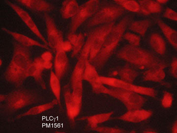
Anti-PLCγ1 (N-terminal region) Antibody
- SPECIFICATION
- CITATIONS
- PROTOCOLS
- BACKGROUND

Application
| WB, IHC |
|---|---|
| Primary Accession | P19174 |
| Reactivity | Bovine, Chicken |
| Host | Mouse |
| Clonality | Mouse Monoclonal |
| Isotype | IgG1 |
| Clone Names | M156 |
| Calculated MW | 148532 Da |
| Gene ID | 5335 |
|---|---|
| Other Names | Phospholipase C gamma1, phosphodiesterase |
| Dilution | WB~~1:1000 IHC~~1:100~500 |
| Storage | Maintain refrigerated at 2-8°C for up to 6 months. For long term storage store at -20°C in small aliquots to prevent freeze-thaw cycles. |
| Precautions | Anti-PLCγ1 (N-terminal region) Antibody is for research use only and not for use in diagnostic or therapeutic procedures. |
| Shipping | Blue Ice |

Thousands of laboratories across the world have published research that depended on the performance of antibodies from Abcepta to advance their research. Check out links to articles that cite our products in major peer-reviewed journals, organized by research category.
info@abcepta.com, and receive a free "I Love Antibodies" mug.

Provided below are standard protocols that you may find useful for product applications.
Background
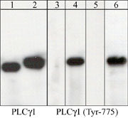
Phosphoinositide-specific phospholipase C (PLC) plays a significant role in transmembrane signaling. In response to extracellular stimuli such as hormones, growth factors, and neurotransmitters, PLC hydrolyzes phosphatidylinositol 4,5-bisphosphate (PIP2) to generate two secondary messengers: inositol 1,4,5-triphosphate (IP3) and diacylglycerol (DAG). At least four families of PLCs have been identified: PLCβ, PLCγ, PLCδ, and PLCε. Phosphorylation is one of the key mechanisms that regulates the activity of PLC. PLCδ is activated by both receptor and nonreceptor tyrosine kinases. PLCγ1 forms a complex with EGF and PDGF receptors, which leads to phosphorylation at tyrosine 771, 783, and 1245. In addition, antigen receptor-induced activation of PLCγ1 leads to phosphorylation at both Tyr-775 and Tyr-783. These two sites are equally important for activation of enzymatic activity.
If you have used an Abcepta product and would like to share how it has performed, please click on the "Submit Review" button and provide the requested information. Our staff will examine and post your review and contact you if needed.
If you have any additional inquiries please email technical services at tech@abcepta.com.
Ordering Information
Other Products
Shipping Information
