


 Foundational characteristics of cancer include proliferation, angiogenesis, migration, evasion of apoptosis, and cellular immortality. Find key markers for these cellular processes and antibodies to detect them.
 The SUMOplot™ Analysis Program predicts and scores sumoylation sites in your protein. SUMOylation is a post-translational modification involved in various cellular processes, such as nuclear-cytosolic transport, transcriptional regulation, apoptosis, protein stability, response to stress, and progression through the cell cycle.
 The Autophagy Receptor Motif Plotter predicts and scores autophagy receptor binding sites in your protein. Identifying proteins connected to this pathway is critical to understanding the role of autophagy in physiological as well as pathological processes such as development, differentiation, neurodegenerative diseases, stress, infection, and cancer.
Anti-Prion Protein (Ser-43), Phosphospecific Antibody
- SPECIFICATION
- CITATIONS
- PROTOCOLS
- BACKGROUND

| Primary Accession | P04156 |
|---|---|
| Reactivity | Bovine |
| Host | Rabbit |
| Clonality | Rabbit Polyclonal |
| Isotype | IgG |
| Calculated MW | 27661 Da |
| Gene ID | 5621 |
|---|---|
| Other Names | PrP, PrPsc, PrPc |
| Storage | Maintain refrigerated at 2-8°C for up to 6 months. For long term storage store at -20°C in small aliquots to prevent freeze-thaw cycles. |
| Precautions | Anti-Prion Protein (Ser-43), Phosphospecific Antibody is for research use only and not for use in diagnostic or therapeutic procedures. |
| Shipping | Blue Ice |

Thousands of laboratories across the world have published research that depended on the performance of antibodies from Abcepta to advance their research. Check out links to articles that cite our products in major peer-reviewed journals, organized by research category.
info@abcepta.com, and receive a free "I Love Antibodies" mug.

Provided below are standard protocols that you may find useful for product applications.
Background
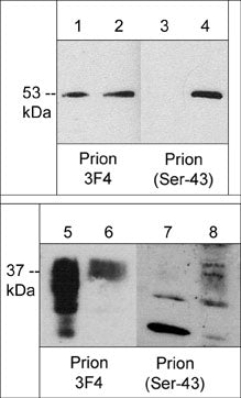
Prion related neurodegenerative diseases, called transmissible spongiform encephalopathies, are observed in many animal species. These diseases involve conversion of normal cellular prion protein (PrPc) into a form that is insoluble and resistant to proteases (PrPSc). The protease resistant form can polymerize into fibrils which accumulate in infected tissues and cause cell death and tissue damage. PrPs have an N-terminal signal sequence and a C-terminal linkage to glycosylphosphatidylinositol anchor. The mature protein is a glycosylated protein that associates with cell membranes. Phosphorylation of PrPC at Ser-43 by Cdk5 promotes proteinase K resistance, prion aggregation, and fibril formation in vitro. In addition, Ser-43 phosphorylation is upregulated in scrapie-infected mouse brain relative to controls. Thus, phosphorylation of Ser-43 may be an important mechanism leading conversion of PrPc to PrPSc and the onset of disease.
If you have used an Abcepta product and would like to share how it has performed, please click on the "Submit Review" button and provide the requested information. Our staff will examine and post your review and contact you if needed.
If you have any additional inquiries please email technical services at tech@abcepta.com.
Ordering Information
Other Products
Shipping Information
