


 Foundational characteristics of cancer include proliferation, angiogenesis, migration, evasion of apoptosis, and cellular immortality. Find key markers for these cellular processes and antibodies to detect them.
 The SUMOplot™ Analysis Program predicts and scores sumoylation sites in your protein. SUMOylation is a post-translational modification involved in various cellular processes, such as nuclear-cytosolic transport, transcriptional regulation, apoptosis, protein stability, response to stress, and progression through the cell cycle.
 The Autophagy Receptor Motif Plotter predicts and scores autophagy receptor binding sites in your protein. Identifying proteins connected to this pathway is critical to understanding the role of autophagy in physiological as well as pathological processes such as development, differentiation, neurodegenerative diseases, stress, infection, and cancer.
Anti-αII-Spectrin , cleavage-specific Antibody
- SPECIFICATION
- CITATIONS
- PROTOCOLS
- BACKGROUND

| Primary Accession | Q13813 |
|---|---|
| Reactivity | Bovine |
| Host | Rabbit |
| Clonality | Rabbit Polyclonal |
| Isotype | IgG |
| Calculated MW | 284539 Da |
| Gene ID | 6709 |
|---|---|
| Other Names | Alpha-II spectrin, Fodrin alpha chain, Spectrin, non-erythroid alpha subunit, SPTAN1, NEAS, SPTA2 |
| Storage | Maintain refrigerated at 2-8°C for up to 6 months. For long term storage store at -20°C in small aliquots to prevent freeze-thaw cycles. |
| Precautions | Anti-αII-Spectrin , cleavage-specific Antibody is for research use only and not for use in diagnostic or therapeutic procedures. |
| Shipping | Blue Ice |

Thousands of laboratories across the world have published research that depended on the performance of antibodies from Abcepta to advance their research. Check out links to articles that cite our products in major peer-reviewed journals, organized by research category.
info@abcepta.com, and receive a free "I Love Antibodies" mug.

Provided below are standard protocols that you may find useful for product applications.
Background
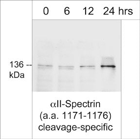
Spectrins are central components of the cytoskeleton that form a scaffold below the plasma membrane. Spectrins contain two subunits, α and β, which intertwine to form heterodimers that can self associate into elongated tetramers. α-spectrin I and β-spectrin I form heterodimers in red blood cells, while nonerythroid mammalian cells contain heterodimers of α-spectrin I and II with β-spectrin I to V. The structure of spectrins includes a succession of triple-helical repeats along with various domains, such as SH3 domain, EF hands, PH domains, and binding domains for ankyrin, actin, band 4.1, and calmodulin. α-spectrin II is a widely expressed non-erythroid spectrin that contains an SH3 domain, a calmodulin binding site, and two cleavage sites, one at Tyr-1176 for calpains and one at Asp-1185 for caspase-3. α-spectrin II and β-spectrin II, like many other spectrins, can form heterodimers that can self associate into tetramers, as well as interact with Band 4.1, F-actin, and other proteins near the plasma membrane.
If you have used an Abcepta product and would like to share how it has performed, please click on the "Submit Review" button and provide the requested information. Our staff will examine and post your review and contact you if needed.
If you have any additional inquiries please email technical services at tech@abcepta.com.
Ordering Information
Other Products
Shipping Information
