


 Foundational characteristics of cancer include proliferation, angiogenesis, migration, evasion of apoptosis, and cellular immortality. Find key markers for these cellular processes and antibodies to detect them.
 The SUMOplot™ Analysis Program predicts and scores sumoylation sites in your protein. SUMOylation is a post-translational modification involved in various cellular processes, such as nuclear-cytosolic transport, transcriptional regulation, apoptosis, protein stability, response to stress, and progression through the cell cycle.
 The Autophagy Receptor Motif Plotter predicts and scores autophagy receptor binding sites in your protein. Identifying proteins connected to this pathway is critical to understanding the role of autophagy in physiological as well as pathological processes such as development, differentiation, neurodegenerative diseases, stress, infection, and cancer.
Anti-VEGFR-2 Antibody
- SPECIFICATION
- CITATIONS
- PROTOCOLS
- BACKGROUND

Application
| WB |
|---|---|
| Primary Accession | P35968 |
| Reactivity | Bovine |
| Host | Rabbit |
| Clonality | Rabbit Polyclonal |
| Isotype | IgG |
| Calculated MW | 151527 Da |
| Gene ID | 3791 |
|---|---|
| Other Names | KDR, flk-1, Vascular endothelial growth factor receptor 2 |
| Dilution | WB~~1:1000 |
| Storage | Maintain refrigerated at 2-8°C for up to 6 months. For long term storage store at -20°C in small aliquots to prevent freeze-thaw cycles. |
| Precautions | Anti-VEGFR-2 Antibody is for research use only and not for use in diagnostic or therapeutic procedures. |
| Shipping | Blue Ice |

Thousands of laboratories across the world have published research that depended on the performance of antibodies from Abcepta to advance their research. Check out links to articles that cite our products in major peer-reviewed journals, organized by research category.
info@abcepta.com, and receive a free "I Love Antibodies" mug.

Provided below are standard protocols that you may find useful for product applications.
Background
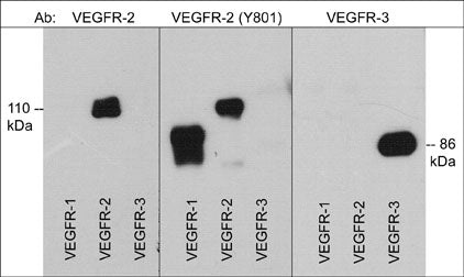
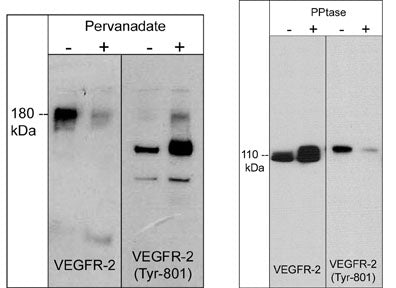
Vascular endothelial growth factor receptor-2 (VEGFR-2/Flk-1/KDR) is the primary receptor for VEGF in endothelial cells. Other VEGFR family members, VEGFR-1 (Flt-1) and VEGFR-3 (Flt-4), can also transduce the intracellular signals of VEGF. However, the role of VEGFR-1 is observed mainly during embryonic angiogenesis and VEGFR-3 signaling may be restricted to specific types of endothelial cells. Major autophosphorylation sites of VEGFR-2 are located in the kinase insert domain (Tyr-951/996) and in the tyrosine kinase catalytic domain (Tyr-1054/1059). Other sites, Tyr-1175 and Tyr-1212 provide docking sites for downstream signaling molecules. Activation of VEGFR-2 also phosphorylates Tyr-801, leading to PI3-kinase-Akt activation and increases in endothelial nitric oxide synthase activity. Phosphorylation of mutliple sites in VEGFR-2 is required for downstream activation of several signaling pathways that control proliferation, chemotaxis, and sprouting during angiogenesis.
If you have used an Abcepta product and would like to share how it has performed, please click on the "Submit Review" button and provide the requested information. Our staff will examine and post your review and contact you if needed.
If you have any additional inquiries please email technical services at tech@abcepta.com.
Ordering Information
Other Products
Shipping Information
