


 Foundational characteristics of cancer include proliferation, angiogenesis, migration, evasion of apoptosis, and cellular immortality. Find key markers for these cellular processes and antibodies to detect them.
 The SUMOplot™ Analysis Program predicts and scores sumoylation sites in your protein. SUMOylation is a post-translational modification involved in various cellular processes, such as nuclear-cytosolic transport, transcriptional regulation, apoptosis, protein stability, response to stress, and progression through the cell cycle.
 The Autophagy Receptor Motif Plotter predicts and scores autophagy receptor binding sites in your protein. Identifying proteins connected to this pathway is critical to understanding the role of autophagy in physiological as well as pathological processes such as development, differentiation, neurodegenerative diseases, stress, infection, and cancer.
MCP-1 Antibody
Purified Mouse Monoclonal Antibody
- SPECIFICATION
- CITATIONS
- PROTOCOLS
- BACKGROUND

Application
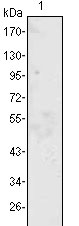
| WB, E |
|---|---|
| Primary Accession | P13500 |
| Reactivity | Human |
| Host | Mouse |
| Clonality | Monoclonal |
| Clone Names | 1A7B8 |
| Isotype | IgG1.kappa |
| Calculated MW | 11025 Da |
| Description | Monocyte chemoattractant protein-1 (MCP-1) is a member of the chemokine-beta family of cytokines. The protein is structurally related to the CXC subfamily of cytokines. Members of this subfamily are characterized by two cysteines separated by a single amino acid. This cytokine displays chemotactic activity for monocytes and basophils but not for neutrophils or eosinophils. It has been implicated in the pathogenesis of diseases characterized by monocytic infiltrates, like psoriasis, rheumatoid arthritis and atherosclerosis. It binds to chemokine receptors CCR2 and CCR4. It may play a inprotant role in the initiation and/or progression of pulmonary hypertension (PH). Blockade of a systemic MCP-1 signal pathway in vivo may prevent PH. |
| Immunogen | Purified recombinant fragment of human MCP-1 expressed in E. Coli. |
| Formulation | Purified antibody in PBS containing 0.03% sodium azide. |
| Gene ID | 6347 |
|---|---|
| Other Names | C-C motif chemokine 2, HC11, Monocyte chemoattractant protein 1, Monocyte chemotactic and activating factor, MCAF, Monocyte chemotactic protein 1, MCP-1, Monocyte secretory protein JE, Small-inducible cytokine A2, CCL2, MCP1, SCYA2 |
| Dilution | WB~~1/500 - 1/2000 E~~N/A |
| Storage | Maintain refrigerated at 2-8°C for up to 6 months. For long term storage store at -20°C in small aliquots to prevent freeze-thaw cycles. |
| Precautions | MCP-1 Antibody is for research use only and not for use in diagnostic or therapeutic procedures. |
| Name | CCL2 |
|---|---|
| Synonyms | MCP1, SCYA2 |
| Function | Acts as a ligand for C-C chemokine receptor CCR2 (PubMed:10529171, PubMed:10587439, PubMed:9837883). Signals through binding and activation of CCR2 and induces a strong chemotactic response and mobilization of intracellular calcium ions (PubMed:10587439, PubMed:9837883). Exhibits a chemotactic activity for monocytes and basophils but not neutrophils or eosinophils (PubMed:8195247, PubMed:8627182, PubMed:9792674). May be involved in the recruitment of monocytes into the arterial wall during the disease process of atherosclerosis (PubMed:8107690). |
| Cellular Location | Secreted |
| Tissue Location | Expressed in the seminal plasma, endometrial fluid and follicular fluid (at protein level) (PubMed:23765988). Expressed in monocytes (PubMed:2513477). |

Thousands of laboratories across the world have published research that depended on the performance of antibodies from Abcepta to advance their research. Check out links to articles that cite our products in major peer-reviewed journals, organized by research category.
info@abcepta.com, and receive a free "I Love Antibodies" mug.

Provided below are standard protocols that you may find useful for product applications.
References
1. Yoshimura T. et al. 1989. FEBS Lett. 244:487-493. 2. Yoshimura T. et al. 1991. Adv. Exp. Med. Biol. 305:47-56. 3. Rollins B.J. et al. 1991. Genomics. 10:489-492.
If you have used an Abcepta product and would like to share how it has performed, please click on the "Submit Review" button and provide the requested information. Our staff will examine and post your review and contact you if needed.
If you have any additional inquiries please email technical services at tech@abcepta.com.
Ordering Information
Other Products
Shipping Information
