


 Foundational characteristics of cancer include proliferation, angiogenesis, migration, evasion of apoptosis, and cellular immortality. Find key markers for these cellular processes and antibodies to detect them.
 The SUMOplot™ Analysis Program predicts and scores sumoylation sites in your protein. SUMOylation is a post-translational modification involved in various cellular processes, such as nuclear-cytosolic transport, transcriptional regulation, apoptosis, protein stability, response to stress, and progression through the cell cycle.
 The Autophagy Receptor Motif Plotter predicts and scores autophagy receptor binding sites in your protein. Identifying proteins connected to this pathway is critical to understanding the role of autophagy in physiological as well as pathological processes such as development, differentiation, neurodegenerative diseases, stress, infection, and cancer.
Calcyclin Antibody
Purified Mouse Monoclonal Antibody
- SPECIFICATION
- CITATIONS
- PROTOCOLS
- BACKGROUND

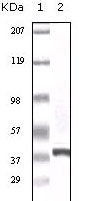
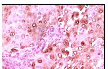
Application
| WB, IHC, E |
|---|---|
| Primary Accession | Q9HB71 |
| Reactivity | Human |
| Host | Mouse |
| Clonality | Monoclonal |
| Clone Names | 7D11 |
| Isotype | IgG1 |
| Calculated MW | 26210 Da |
| Description | Calcyclin encoded by this gene is a member of the S100 family of proteins containing 2 EF-hand calcium-binding motifs. S100 proteins are localized in the cytoplasm and/or nucleus of a wide range of cells, and involved in the regulation of a number of cellular processes such as cell cycle progression and differentiation. S100 genes include at least 13 members which are located as a cluster on chromosome 1q21. This protein may function in stimulation of Ca2+-dependent insulin release, stimulation of prolactin secretion, and exocytosis. Chromosomal rearrangements and altered expression of this gene have been implicated in melanoma. |
| Immunogen | Purified recombinant fragment of calcyclin expressed in E. Coli. |
| Formulation | Purified antibody in PBS containing 0.03% sodium azide. |
| Gene ID | 27101 |
|---|---|
| Other Names | Calcyclin-binding protein, CacyBP, hCacyBP, S100A6-binding protein, Siah-interacting protein, CACYBP, S100A6BP, SIP |
| Dilution | WB~~1/500 - 1/2000 IHC~~1/200 - 1/1000 E~~N/A |
| Storage | Maintain refrigerated at 2-8°C for up to 6 months. For long term storage store at -20°C in small aliquots to prevent freeze-thaw cycles. |
| Precautions | Calcyclin Antibody is for research use only and not for use in diagnostic or therapeutic procedures. |
| Name | CACYBP |
|---|---|
| Synonyms | S100A6BP, SIP |
| Function | May be involved in calcium-dependent ubiquitination and subsequent proteasomal degradation of target proteins. Probably serves as a molecular bridge in ubiquitin E3 complexes. Participates in the ubiquitin-mediated degradation of beta-catenin (CTNNB1). |
| Cellular Location | Nucleus. Cytoplasm. Note=Cytoplasmic at low calcium concentrations. In neuroblastoma cells, after a retinoic acid (RA) induction and calcium increase, it localizes in both the nucleus and cytoplasm. The nuclear fraction may be phosphorylated |

Thousands of laboratories across the world have published research that depended on the performance of antibodies from Abcepta to advance their research. Check out links to articles that cite our products in major peer-reviewed journals, organized by research category.
info@abcepta.com, and receive a free "I Love Antibodies" mug.

Provided below are standard protocols that you may find useful for product applications.
References
1. Bean C.et.al J Mol Biol. 2005 Jun 3;349(2):349-66. 2. HWang R.et.al J Biol Chem. 2004 May 14;279(20):21239-47.
If you have used an Abcepta product and would like to share how it has performed, please click on the "Submit Review" button and provide the requested information. Our staff will examine and post your review and contact you if needed.
If you have any additional inquiries please email technical services at tech@abcepta.com.
Ordering Information
Other Products
Shipping Information
