


 Foundational characteristics of cancer include proliferation, angiogenesis, migration, evasion of apoptosis, and cellular immortality. Find key markers for these cellular processes and antibodies to detect them.
 The SUMOplot™ Analysis Program predicts and scores sumoylation sites in your protein. SUMOylation is a post-translational modification involved in various cellular processes, such as nuclear-cytosolic transport, transcriptional regulation, apoptosis, protein stability, response to stress, and progression through the cell cycle.
 The Autophagy Receptor Motif Plotter predicts and scores autophagy receptor binding sites in your protein. Identifying proteins connected to this pathway is critical to understanding the role of autophagy in physiological as well as pathological processes such as development, differentiation, neurodegenerative diseases, stress, infection, and cancer.
EphB6 Antibody
Purified Mouse Monoclonal Antibody
- SPECIFICATION
- CITATIONS
- PROTOCOLS
- BACKGROUND

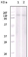
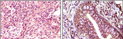
Application
| WB, IHC, E |
|---|---|
| Primary Accession | O15197 |
| Reactivity | Human |
| Host | Mouse |
| Clonality | Monoclonal |
| Clone Names | 8E7H12 |
| Isotype | IgG1 |
| Calculated MW | 110700 Da |
| Description | EPH receptor B6 (EphB6), with 1006-amino acid protein(~ 110 kDa), belongs to the ephrin receptor subfamily of the protein-tyrosine kinase family. Eph receptors and ephrin ligands are membrane-bound cell-cell communication molecules with well-defined functions in development. EphB6 is expressed both in a variety of embryonic and adult tissues. EphB6 is a unique member in the Eph family of receptor tyrosine kinases in that its kinase domain contains several alterations in conserved amino acids and is catalytically inactive. EphB6 can both positively and negatively regulate cell adhesion and migration and tyrosine phosphorylation of the receptor by an Src family kinase acts as the molecular switch for the functional transition. |
| Immunogen | Purified recombinant fragment of EphB6 expressed in E. Coli. |
| Formulation | Purified antibody in PBS containing 0.03% sodium azide. |
| Gene ID | 2051 |
|---|---|
| Other Names | Ephrin type-B receptor 6, HEP, Tyrosine-protein kinase-defective receptor EPH-6, EPHB6 |
| Dilution | WB~~1/500 - 1/2000 IHC~~1/200 - 1/1000 E~~N/A |
| Storage | Maintain refrigerated at 2-8°C for up to 6 months. For long term storage store at -20°C in small aliquots to prevent freeze-thaw cycles. |
| Precautions | EphB6 Antibody is for research use only and not for use in diagnostic or therapeutic procedures. |
| Name | EPHB6 |
|---|---|
| Function | Kinase-defective receptor for members of the ephrin-B family. Binds to ephrin-B1 and ephrin-B2. Modulates cell adhesion and migration by exerting both positive and negative effects upon stimulation with ephrin-B2. Inhibits JNK activation, T-cell receptor-induced IL-2 secretion and CD25 expression upon stimulation with ephrin-B2. |
| Cellular Location | Membrane; Single-pass type I membrane protein. |
| Tissue Location | Expressed in brain. Expressed in non invasive breast carcinoma cell lines (at protein level). Strong expression in brain and pancreas, and weak expression in other tissues, such as heart, placenta, lung, liver, skeletal muscle and kidney. Expressed in breast non invasive tumors but not in metastatic lesions. Isoform 3 is expressed in cell lines of glioblastomas, anaplastic astrocytomas, gliosarcomas and astrocytomas. Isoform 3 is not detected in normal tissues. |

Thousands of laboratories across the world have published research that depended on the performance of antibodies from Abcepta to advance their research. Check out links to articles that cite our products in major peer-reviewed journals, organized by research category.
info@abcepta.com, and receive a free "I Love Antibodies" mug.

Provided below are standard protocols that you may find useful for product applications.
References
1. Kazushige Ogawa, Hiroki Wada, Noriyoshi Okada J Cell Sci. 2006 Feb 1;119(Pt 3):559-70. 2. Hiroshi Matsuoka, Hiroya Obama, Meghan L. Kelly J Biol Chem. 2005 Aug 12;280(32):29355-63.
If you have used an Abcepta product and would like to share how it has performed, please click on the "Submit Review" button and provide the requested information. Our staff will examine and post your review and contact you if needed.
If you have any additional inquiries please email technical services at tech@abcepta.com.
Ordering Information
Other Products
Shipping Information
