


 Foundational characteristics of cancer include proliferation, angiogenesis, migration, evasion of apoptosis, and cellular immortality. Find key markers for these cellular processes and antibodies to detect them.
 The SUMOplot™ Analysis Program predicts and scores sumoylation sites in your protein. SUMOylation is a post-translational modification involved in various cellular processes, such as nuclear-cytosolic transport, transcriptional regulation, apoptosis, protein stability, response to stress, and progression through the cell cycle.
 The Autophagy Receptor Motif Plotter predicts and scores autophagy receptor binding sites in your protein. Identifying proteins connected to this pathway is critical to understanding the role of autophagy in physiological as well as pathological processes such as development, differentiation, neurodegenerative diseases, stress, infection, and cancer.
CIB1 Antibody
Purified Mouse Monoclonal Antibody
- SPECIFICATION
- CITATIONS
- PROTOCOLS
- BACKGROUND

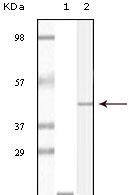
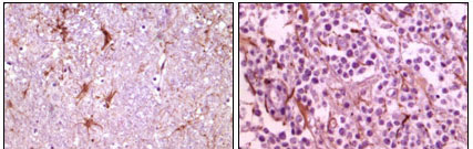
Application
| WB, IHC, E |
|---|---|
| Primary Accession | Q99828 |
| Reactivity | Human |
| Host | Mouse |
| Clonality | Monoclonal |
| Clone Names | 5A1F5E12; 5A1H7E12 |
| Calculated MW | 21703 Da |
| Description | CIB1(also designated calcium and integrin binding 1 or calmyrin),with 191-amino acid protein(about 21kDa), belongs to the calcium-binding protein family.CIB1 is known to interact with DNA-dependent protein kinase and may play a role in kinase-phosphatase regulation of DNA end joining.CIB1 is an EF-hand-containing protein that binds multiple effector proteins, including the platelet alpha(IIb)beta(3) integrin and several serine/threonine kinases and potentially modulates their function.CIB1 regulates platelet aggregation in hemostasis through a specific interaction with the alpha(IIb) cytoplasmic domain of platelet integrin alpha(IIb)beta(3). CIB1 is also ubiquitously expressed activating and inhibiting protein ligand of the InsP3R. |
| Immunogen | Purified recombinant fragment of CIB1 expressed in E. Coli. |
| Formulation | Ascitic fluid containing 0.03% sodium azide. |
| Gene ID | 10519 |
|---|---|
| Other Names | Calcium and integrin-binding protein 1, CIB, Calcium- and integrin-binding protein, CIBP, Calmyrin, DNA-PKcs-interacting protein, Kinase-interacting protein, KIP, SNK-interacting protein 2-28, SIP2-28, CIB1, CIB, KIP, PRKDCIP |
| Dilution | WB~~1/500 - 1/2000 IHC~~1/200 - 1/1000 E~~N/A |
| Storage | Maintain refrigerated at 2-8°C for up to 6 months. For long term storage store at -20°C in small aliquots to prevent freeze-thaw cycles. |
| Precautions | CIB1 Antibody is for research use only and not for use in diagnostic or therapeutic procedures. |
| Name | CIB1 |
|---|---|
| Synonyms | CIB, KIP, PRKDCIP |
| Function | Calcium-binding protein that plays a role in the regulation of numerous cellular processes, such as cell differentiation, cell division, cell proliferation, cell migration, thrombosis, angiogenesis, cardiac hypertrophy and apoptosis. Involved in bone marrow megakaryocyte differentiation by negatively regulating thrombopoietin- mediated signaling pathway. Participates in the endomitotic cell cycle of megakaryocyte, a form of mitosis in which both karyokinesis and cytokinesis are interrupted. Plays a role in integrin signaling by negatively regulating alpha-IIb/beta3 activation in thrombin-stimulated megakaryocytes preventing platelet aggregation. Up-regulates PTK2/FAK1 activity, and is also needed for the recruitment of PTK2/FAK1 to focal adhesions; it thus appears to play an important role in focal adhesion formation. Positively regulates cell migration on fibronectin in a CDC42-dependent manner, the effect being negatively regulated by PAK1. Functions as a negative regulator of stress activated MAP kinase (MAPK) signaling pathways. Down-regulates inositol 1,4,5-trisphosphate receptor-dependent calcium signaling. Involved in sphingosine kinase SPHK1 translocation to the plasma membrane in a N-myristoylation- dependent manner preventing TNF-alpha-induced apoptosis. Regulates serine/threonine-protein kinase PLK3 activity for proper completion of cell division progression. Plays a role in microtubule (MT) dynamics during neuronal development; disrupts the MT depolymerization activity of STMN2 attenuating NGF-induced neurite outgrowth and the MT reorganization at the edge of lamellipodia. Promotes cardiomyocyte hypertrophy via activation of the calcineurin/NFAT signaling pathway. Stimulates calcineurin PPP3R1 activity by mediating its anchoring to the sarcolemma. In ischemia-induced (pathological or adaptive) angiogenesis, stimulates endothelial cell proliferation, migration and microvessel formation by activating the PAK1 and ERK1/ERK2 signaling pathway. Also promotes cancer cell survival and proliferation. May regulate cell cycle and differentiation of spermatogenic germ cells, and/or differentiation of supporting Sertoli cells. Forms a complex with TMC6/EVER1 and TMC8/EVER2 in lymphocytes and keratynocytes where CIB1 stabilizes TMC6 and TMC8 levels and reciprocally (PubMed:30068544, PubMed:32917726). |
| Cellular Location | Membrane; Lipid-anchor. Cell membrane, sarcolemma. Cell membrane. Apical cell membrane. Cell projection, ruffle membrane. Cell projection, filopodium tip. Cell projection, growth cone. Cell projection, lamellipodium. Cytoplasm. Cytoplasm, cytoskeleton. Cytoplasm, cytoskeleton, microtubule organizing center, centrosome. Cytoplasm, perinuclear region. Nucleus. Cell projection, neuron projection. Perikaryon. Note=Colocalized with PPP3R1 at the cell membrane of cardiomyocytes in the hypertrophic heart (By similarity) Colocalized with NBR1 to the perinuclear region. Colocalizes with TAS1R2 in apical regions of taste receptor cells. Colocalized with RAC3 in the perinuclear area and at the cell periphery. Colocalized with PAK1 within membrane ruffles during cell spreading upon readhesion to fibronectin. Redistributed to the cytoskeleton upon platelet aggregation. Translocates from the cytosol to the plasma membrane in a calcium-dependent manner. Colocalized with PLK3 at centrosomes in ductal breast carcinoma cells. |
| Tissue Location | Ubiquitously expressed. Expressed in the epidermis, hair follicles and keratinocytes (PubMed:30068544). Detected in platelets and in cell lines of megakaryocytic and erythrocytic lineages. Both isoform 1 and isoform 2 are detected in various cancer cell lines, with isoform 2 being the predominant form (at protein level). |

Thousands of laboratories across the world have published research that depended on the performance of antibodies from Abcepta to advance their research. Check out links to articles that cite our products in major peer-reviewed journals, organized by research category.
info@abcepta.com, and receive a free "I Love Antibodies" mug.

Provided below are standard protocols that you may find useful for product applications.
References
1. Holly R. Gentry,Alex U. Singer, Laurie Betts. J. Biol. Chem., Mar 2005; 280: 8407 - 8415. 2. Carl White, Jun Yang, Mervyn J. Monteiro. J. Biol. Chem., Jul 2006; 281: 20825 – 20833.
If you have used an Abcepta product and would like to share how it has performed, please click on the "Submit Review" button and provide the requested information. Our staff will examine and post your review and contact you if needed.
If you have any additional inquiries please email technical services at tech@abcepta.com.
Ordering Information
Other Products
Shipping Information
