


 Foundational characteristics of cancer include proliferation, angiogenesis, migration, evasion of apoptosis, and cellular immortality. Find key markers for these cellular processes and antibodies to detect them.
 The SUMOplot™ Analysis Program predicts and scores sumoylation sites in your protein. SUMOylation is a post-translational modification involved in various cellular processes, such as nuclear-cytosolic transport, transcriptional regulation, apoptosis, protein stability, response to stress, and progression through the cell cycle.
 The Autophagy Receptor Motif Plotter predicts and scores autophagy receptor binding sites in your protein. Identifying proteins connected to this pathway is critical to understanding the role of autophagy in physiological as well as pathological processes such as development, differentiation, neurodegenerative diseases, stress, infection, and cancer.
ELK1 Antibody
Purified Mouse Monoclonal Antibody
- SPECIFICATION
- CITATIONS
- PROTOCOLS
- BACKGROUND

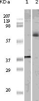
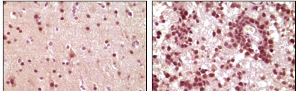
Application
| WB, IHC, E |
|---|---|
| Primary Accession | P19419 |
| Reactivity | Human |
| Host | Mouse |
| Clonality | Monoclonal |
| Clone Names | 3H6D12; 4H9C8; 4H9F1 |
| Isotype | IgG1 |
| Calculated MW | 44888 Da |
| Description | The transcription factor ELK1 is a family of member of ETS oncogene family and of the ternary complex factor (TCF) subfamily,which is located on chromosome Xp11.2 and stimulates transcription. binds to purine-rich DNA sequences. Proteins of the TCF subfamily form a ternary complex by binding to the the serum response factor and the serum reponse element in the promoter of the c-fos proto-oncogene. The protein encoded by this gene is a nuclear target for the ras-raf-MAPK signaling cascade. Elk1 is phosphorylated by MAP kinase pathways at a cluster of S/T motifs at its C terminus,It appears to be a direct target of activated MAP kinase. Biochemical studies indicate that Elk1 is a good substrate for MAP kinase, the kinetics of Elk1phosphorylation and activation correlate with MAP kinase activity, and interfering mutants of MAP kinase block Elk1 activation in vivo. More recent studies have shown that Elk1 is also a target of the Stress Activated Kinase SAPK/JNK. Phosphorylation of Elk1 has also been implicated in synaptic plasticity in the adult hippocampus. |
| Immunogen | Purified recombinant fragment of ELK1 expressed in E. Coli. |
| Formulation | Ascitic fluid containing 0.03% sodium azide. |
| Gene ID | 2002 |
|---|---|
| Other Names | ETS domain-containing protein Elk-1, ELK1 |
| Dilution | WB~~1/500 - 1/2000 IHC~~1/200 - 1/1000 E~~N/A |
| Storage | Maintain refrigerated at 2-8°C for up to 6 months. For long term storage store at -20°C in small aliquots to prevent freeze-thaw cycles. |
| Precautions | ELK1 Antibody is for research use only and not for use in diagnostic or therapeutic procedures. |
| Name | ELK1 (HGNC:3321) |
|---|---|
| Function | Transcription factor that binds to purine-rich DNA sequences (PubMed:10799319, PubMed:7889942). Forms a ternary complex with SRF and the ETS and SRF motifs of the serum response element (SRE) on the promoter region of immediate early genes such as FOS and IER2 (PubMed:1630903). Induces target gene transcription upon JNK and MAPK- signaling pathways stimulation (PubMed:7889942). |
| Cellular Location | Nucleus. |
| Tissue Location | Lung and testis. |

Thousands of laboratories across the world have published research that depended on the performance of antibodies from Abcepta to advance their research. Check out links to articles that cite our products in major peer-reviewed journals, organized by research category.
info@abcepta.com, and receive a free "I Love Antibodies" mug.

Provided below are standard protocols that you may find useful for product applications.
References
1. Rao,V.N., et al. 1989.Science.244 (4900):66-70. 2. Hsieh,Y.H., et al. 2006.Biochem. Biophys. Res. Commun. 339 (1): 217-225. 3. Gille,H., Strahl,T. and Shaw,P.E.1995. Curr. Biol. 5 (10): 1191-1200. 4. Gille,H., et al. 1995.EMBO J. 14 (5): 951-962.
If you have used an Abcepta product and would like to share how it has performed, please click on the "Submit Review" button and provide the requested information. Our staff will examine and post your review and contact you if needed.
If you have any additional inquiries please email technical services at tech@abcepta.com.
Ordering Information
Other Products
Shipping Information
