


 Foundational characteristics of cancer include proliferation, angiogenesis, migration, evasion of apoptosis, and cellular immortality. Find key markers for these cellular processes and antibodies to detect them.
 The SUMOplot™ Analysis Program predicts and scores sumoylation sites in your protein. SUMOylation is a post-translational modification involved in various cellular processes, such as nuclear-cytosolic transport, transcriptional regulation, apoptosis, protein stability, response to stress, and progression through the cell cycle.
 The Autophagy Receptor Motif Plotter predicts and scores autophagy receptor binding sites in your protein. Identifying proteins connected to this pathway is critical to understanding the role of autophagy in physiological as well as pathological processes such as development, differentiation, neurodegenerative diseases, stress, infection, and cancer.
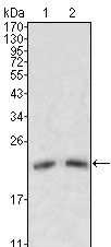
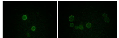
ApoM Antibody
Purified Mouse Monoclonal Antibody
- SPECIFICATION
- CITATIONS
- PROTOCOLS
- BACKGROUND

Application
| WB, ICC, E |
|---|---|
| Primary Accession | O95445 |
| Reactivity | Human |
| Host | Mouse |
| Clonality | Monoclonal |
| Clone Names | 8F12C6B8 |
| Isotype | IgG1 |
| Calculated MW | 21kDa |
| Description | ApoM (apolipoprotein M, also designated G3a or NG20), with 188-amino acid protein(about 21kDa), is an apolipoprotein and member of the lipocalin protein family. The Apo-proteins are involved in the specific binding of cellular receptors, the regulation of lipolytic enzymes, and the process of lipid exchange. The encoded protein is secreted through the plasma membrane but remains membrane-bound, where it is involved in lipid transport. The N-terminal region of Apo-M contains hydrophobic residues that may promote association with the phospholipid layer of lipoprotein particles. In vitro, Apo-M is glycosylated when translated in the presence of microsomes, and remains associated with the microsomes after carbonate treatment. Apo-M is expressed in liver and kidney, and is secreted into the bloodstream in HDLs, and also found in triglyceride-rich lipoproteins and LDLs. |
| Immunogen | Purified recombinant fragment of human ApoM expressed in E. Coli. |
| Formulation | Purified antibody in PBS containing 0.03% sodium azide. |
| Gene ID | 55937 |
|---|---|
| Other Names | Apolipoprotein M, Apo-M, ApoM, Protein G3a, APOM, G3A, NG20 |
| Dilution | WB~~1/500 - 1/2000 ICC~~N/A E~~N/A |
| Storage | Maintain refrigerated at 2-8°C for up to 6 months. For long term storage store at -20°C in small aliquots to prevent freeze-thaw cycles. |
| Precautions | ApoM Antibody is for research use only and not for use in diagnostic or therapeutic procedures. |
| Name | APOM |
|---|---|
| Synonyms | G3A, NG20 |
| Function | Probably involved in lipid transport. Can bind sphingosine-1- phosphate, myristic acid, palmitic acid and stearic acid, retinol, all- trans-retinoic acid and 9-cis-retinoic acid. |
| Cellular Location | Secreted. Note=Present in high density lipoprotein (HDL) and to a lesser extent in triglyceride-rich lipoproteins (TGRLP) and low density lipoproteins (LDL) |
| Tissue Location | Plasma protein. Expressed in liver and kidney. |

Thousands of laboratories across the world have published research that depended on the performance of antibodies from Abcepta to advance their research. Check out links to articles that cite our products in major peer-reviewed journals, organized by research category.
info@abcepta.com, and receive a free "I Love Antibodies" mug.

Provided below are standard protocols that you may find useful for product applications.
References
1. Xu, N. & Dahlback, B. 1999 J. Biol. Chem. 274:31286 2. Duan J, Dahlback B, Villoutreix BO.FEBS Lett. 2001 Jun 15;499(1-2):127-32. 3. Xu,N., Nilsson-Ehle,P. & Ahren,B. 2004. J. Nutr.Biochem. 15 (10):579-582 4. Zhang,X.Y. ,et al.2004. Acta Histochem. 106 (2):123-128
If you have used an Abcepta product and would like to share how it has performed, please click on the "Submit Review" button and provide the requested information. Our staff will examine and post your review and contact you if needed.
If you have any additional inquiries please email technical services at tech@abcepta.com.
Ordering Information
Other Products
Shipping Information
