


 Foundational characteristics of cancer include proliferation, angiogenesis, migration, evasion of apoptosis, and cellular immortality. Find key markers for these cellular processes and antibodies to detect them.
 The SUMOplot™ Analysis Program predicts and scores sumoylation sites in your protein. SUMOylation is a post-translational modification involved in various cellular processes, such as nuclear-cytosolic transport, transcriptional regulation, apoptosis, protein stability, response to stress, and progression through the cell cycle.
 The Autophagy Receptor Motif Plotter predicts and scores autophagy receptor binding sites in your protein. Identifying proteins connected to this pathway is critical to understanding the role of autophagy in physiological as well as pathological processes such as development, differentiation, neurodegenerative diseases, stress, infection, and cancer.
FER Antibody
Purified Mouse Monoclonal Antibody
- SPECIFICATION
- CITATIONS
- PROTOCOLS
- BACKGROUND

Application
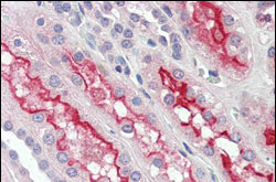
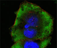
| WB, IHC, ICC, E |
|---|---|
| Primary Accession | P16591 |
| Reactivity | Human, Mouse |
| Host | Mouse |
| Clonality | Monoclonal |
| Clone Names | 5D2C4 |
| Isotype | IgG1 |
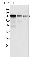
| Calculated MW | 95kDa |
| Description | FER (fer tyrosine kinase) is a member of the FPS/FES family of nontransmembrane receptor tyrosine kinases, which shares a functional domain and is involved in signaling pathways through receptor tyrosine kinases (RTK) and cytokine receptors. The Fes /Fps family is distinct from c-Src, c-Abl and related nRTKs and was originally distinguished as a homolog to retroviral oncoproteins. In vivo, Fer kinase assembles into homotrimers via conserved coiled-coil domains. The N-terminal coiled-coil domains of Fer can autophosphorylate in trans, thereby regulating their cellular function through differential phosphorylation states. Growth factor exposure can induce tyrosine phosphorylation of Fer and recruitment of Fer to RTK complexes containing p85. It is expressed predominantly in mature hematopoietic cells of the granulocytic and monocytic lineage, and has been shown to be expressed in vascular endothelial cells. Fer is implicated in insulin signaling, cell-cell signaling, human prostatic proliferative diseases, and is involved in the regulation of G1 progression. |
| Immunogen | Purified recombinant fragment of human FER expressed in E. Coli. |
| Formulation | Ascitic fluid containing 0.03% sodium azide. |
| Gene ID | 2241 |
|---|---|
| Other Names | Tyrosine-protein kinase Fer, 2.7.10.2, Feline encephalitis virus-related kinase FER, Fujinami poultry sarcoma/Feline sarcoma-related protein Fer, Proto-oncogene c-Fer, Tyrosine kinase 3, p94-Fer, FER, TYK3 |
| Dilution | WB~~1/500 - 1/2000 IHC~~1/200 - 1/1000 ICC~~N/A E~~N/A |
| Storage | Maintain refrigerated at 2-8°C for up to 6 months. For long term storage store at -20°C in small aliquots to prevent freeze-thaw cycles. |
| Precautions | FER Antibody is for research use only and not for use in diagnostic or therapeutic procedures. |
| Name | FER |
|---|---|
| Synonyms | TYK3 |
| Function | Tyrosine-protein kinase that acts downstream of cell surface receptors for growth factors and plays a role in the regulation of the actin cytoskeleton, microtubule assembly, lamellipodia formation, cell adhesion, cell migration and chemotaxis. Acts downstream of EGFR, KIT, PDGFRA and PDGFRB. Acts downstream of EGFR to promote activation of NF- kappa-B and cell proliferation. May play a role in the regulation of the mitotic cell cycle. Plays a role in the insulin receptor signaling pathway and in activation of phosphatidylinositol 3-kinase. Acts downstream of the activated FCER1 receptor and plays a role in FCER1 (high affinity immunoglobulin epsilon receptor)-mediated signaling in mast cells. Plays a role in the regulation of mast cell degranulation. Plays a role in leukocyte recruitment and diapedesis in response to bacterial lipopolysaccharide (LPS). Plays a role in synapse organization, trafficking of synaptic vesicles, the generation of excitatory postsynaptic currents and neuron-neuron synaptic transmission. Plays a role in neuronal cell death after brain damage. Phosphorylates CTTN, CTNND1, PTK2/FAK1, GAB1, PECAM1 and PTPN11. May phosphorylate JUP and PTPN1. Can phosphorylate STAT3, but the biological relevance of this depends on cell type and stimulus. |
| Cellular Location | Cytoplasm. Cytoplasm, cytoskeleton. Cell membrane; Peripheral membrane protein; Cytoplasmic side. Cell projection. Cell junction. Membrane; Peripheral membrane protein; Cytoplasmic side. Nucleus. Cytoplasm, cell cortex. Note=Associated with the chromatin. Detected on microtubules in polarized and motile vascular endothelial cells. Colocalizes with F-actin at the cell cortex. Colocalizes with PECAM1 and CTNND1 at nascent cell-cell contacts |
| Tissue Location | Isoform 1 is detected in normal colon and in fibroblasts (at protein level). Isoform 3 is detected in normal testis, in colon carcinoma-derived metastases in lung, liver and ovary, and in colon carcinoma and hepato carcinoma cell lines (at protein level) Isoform 3 is not detected in normal colon or in normal fibroblasts (at protein level). Widely expressed. |

Thousands of laboratories across the world have published research that depended on the performance of antibodies from Abcepta to advance their research. Check out links to articles that cite our products in major peer-reviewed journals, organized by research category.
info@abcepta.com, and receive a free "I Love Antibodies" mug.

Provided below are standard protocols that you may find useful for product applications.
References
1. Li L. Cheng X. Ling HQ. Plant Mol Biol. 2004, Jan, 54(1):125-36. 2. Girault JA. Greengard P. Arch Neurol. 2004, May, 61(5):641-4. 3. Fan L. Di Ciano-Oliveira C. Weed SA. et al. Biochem J. 2004, Jun 1, 380(Pt 2):581-91.
If you have used an Abcepta product and would like to share how it has performed, please click on the "Submit Review" button and provide the requested information. Our staff will examine and post your review and contact you if needed.
If you have any additional inquiries please email technical services at tech@abcepta.com.
Ordering Information
Other Products
Shipping Information
