


 Foundational characteristics of cancer include proliferation, angiogenesis, migration, evasion of apoptosis, and cellular immortality. Find key markers for these cellular processes and antibodies to detect them.
 The SUMOplot™ Analysis Program predicts and scores sumoylation sites in your protein. SUMOylation is a post-translational modification involved in various cellular processes, such as nuclear-cytosolic transport, transcriptional regulation, apoptosis, protein stability, response to stress, and progression through the cell cycle.
 The Autophagy Receptor Motif Plotter predicts and scores autophagy receptor binding sites in your protein. Identifying proteins connected to this pathway is critical to understanding the role of autophagy in physiological as well as pathological processes such as development, differentiation, neurodegenerative diseases, stress, infection, and cancer.
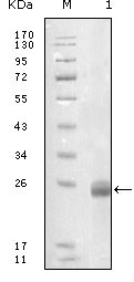
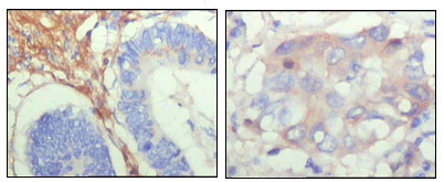
FBLN5 Antibody
Purified Mouse Monoclonal Antibody
- SPECIFICATION
- CITATIONS
- PROTOCOLS
- BACKGROUND

Application
| WB, IHC, E |
|---|---|
| Primary Accession | Q9UBX5 |
| Reactivity | Human |
| Host | Mouse |
| Clonality | Monoclonal |
| Clone Names | 1G6A4 |
| Isotype | IgG1 |
| Calculated MW | 50180 Da |
| Description | FBLN5: fibulin 5. The protein is a secreted, extracellular matrix protein containing an Arg-Gly-Asp (RGD) motif and calcium-binding EGF-like domains. It promotes adhesion of endothelial cells through interaction of integrins and the RGD motif. It is prominently expressed in developing arteries but less so in adult vessels. However, its expression is reinduced in balloon-injured vessels and atherosclerotic lesions, notably in intimal vascular smooth muscle cells and endothelial cells. Therefore, the protein encoded by this gene may play a role in vascular development and remodeling. |
| Immunogen | Purified recombinant fragment of FBLN5 (aa242-448) expressed in E. Coli. |
| Formulation | Ascitic fluid containing 0.03% sodium azide. |
| Gene ID | 10516 |
|---|---|
| Other Names | Fibulin-5, FIBL-5, Developmental arteries and neural crest EGF-like protein, Dance, Urine p50 protein, UP50, FBLN5, DANCE |
| Dilution | WB~~1/500 - 1/2000 IHC~~1/200 - 1/1000 E~~N/A |
| Storage | Maintain refrigerated at 2-8°C for up to 6 months. For long term storage store at -20°C in small aliquots to prevent freeze-thaw cycles. |
| Precautions | FBLN5 Antibody is for research use only and not for use in diagnostic or therapeutic procedures. |
| Name | FBLN5 |
|---|---|
| Synonyms | DANCE |
| Function | Essential for elastic fiber formation, is involved in the assembly of continuous elastin (ELN) polymer and promotes the interaction of microfibrils and ELN (PubMed:18185537). Stabilizes and organizes elastic fibers in the skin, lung and vasculature (By similarity). Promotes adhesion of endothelial cells through interaction of integrins and the RGD motif. Vascular ligand for integrin receptors which may play a role in vascular development and remodeling (PubMed:10428823). May act as an adapter that mediates the interaction between FBN1 and ELN (PubMed:17255108). |
| Cellular Location | Secreted. Secreted, extracellular space, extracellular matrix. Note=co-localizes with ELN in elastic fibers. |
| Tissue Location | Expressed in skin fibroblasts (at protein level) (PubMed:17035250). Expressed predominantly in heart, ovary, and colon but also in kidney, pancreas, testis, lung and placenta. Not detectable in brain, liver, thymus, prostate, or peripheral blood leukocytes (PubMed:10428823). |

Thousands of laboratories across the world have published research that depended on the performance of antibodies from Abcepta to advance their research. Check out links to articles that cite our products in major peer-reviewed journals, organized by research category.
info@abcepta.com, and receive a free "I Love Antibodies" mug.

Provided below are standard protocols that you may find useful for product applications.
References
1. Am J Hum Genet. 2003 Apr;72(4):998-1004. 2. Genome Res. 2003 Oct;13(10):2265-70 3. N Engl J Med. 2004 Jul 22;351(4):346-53. 4. Surgery. 2005 Aug;138(2):352-9.
If you have used an Abcepta product and would like to share how it has performed, please click on the "Submit Review" button and provide the requested information. Our staff will examine and post your review and contact you if needed.
If you have any additional inquiries please email technical services at tech@abcepta.com.
Ordering Information
Other Products
Shipping Information
