


 Foundational characteristics of cancer include proliferation, angiogenesis, migration, evasion of apoptosis, and cellular immortality. Find key markers for these cellular processes and antibodies to detect them.
 The SUMOplot™ Analysis Program predicts and scores sumoylation sites in your protein. SUMOylation is a post-translational modification involved in various cellular processes, such as nuclear-cytosolic transport, transcriptional regulation, apoptosis, protein stability, response to stress, and progression through the cell cycle.
 The Autophagy Receptor Motif Plotter predicts and scores autophagy receptor binding sites in your protein. Identifying proteins connected to this pathway is critical to understanding the role of autophagy in physiological as well as pathological processes such as development, differentiation, neurodegenerative diseases, stress, infection, and cancer.
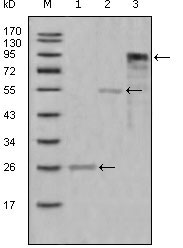
Myeloperoxidase Antibody
Purified Mouse Monoclonal Antibody
- SPECIFICATION
- CITATIONS
- PROTOCOLS
- BACKGROUND

Application
| WB, E |
|---|---|
| Primary Accession | P05164 |
| Reactivity | Human |
| Host | Mouse |
| Clonality | Monoclonal |
| Clone Names | 9B12G7; 4D8B12; 9B12D9; 9C11A5 |
| Isotype | IgG1 |
| Calculated MW | 83869 Da |
| Description | Myeloperoxidase (MPO) is a heme protein synthesized during myeloid differentiation that constitutes the major component of neutrophil azurophilic granules. Produced as a single chain precursor, myeloperoxidase is subsequently cleaved into a light and heavy chain. The mature myeloperoxidase is a tetramer composed of 2 light chains and 2 heavy chains. This enzyme produces hypohalous acids central to the microbicidal activity of netrophils. |
| Immunogen | Purified recombinant fragment of MPO (aa1-193) expressed in E. Coli. |
| Formulation | Ascitic fluid containing 0.03% sodium azide. |
| Gene ID | 4353 |
|---|---|
| Other Names | Myeloperoxidase, MPO, 1.11.2.2, Myeloperoxidase, 89 kDa myeloperoxidase, 84 kDa myeloperoxidase, Myeloperoxidase light chain, Myeloperoxidase heavy chain, MPO |
| Dilution | WB~~1/500 - 1/2000 E~~N/A |
| Storage | Maintain refrigerated at 2-8°C for up to 6 months. For long term storage store at -20°C in small aliquots to prevent freeze-thaw cycles. |
| Precautions | Myeloperoxidase Antibody is for research use only and not for use in diagnostic or therapeutic procedures. |
| Name | MPO (HGNC:7218) |
|---|---|
| Function | Part of the host defense system of polymorphonuclear leukocytes. It is responsible for microbicidal activity against a wide range of organisms. In the stimulated PMN, MPO catalyzes the production of hypohalous acids, primarily hypochlorous acid in physiologic situations, and other toxic intermediates that greatly enhance PMN microbicidal activity (PubMed:9922160). Mediates the proteolytic cleavage of alpha-1-microglobulin to form t-alpha-1-microglobulin, which potently inhibits oxidation of low-density lipoprotein particles and limits vascular damage (PubMed:25698971). |
| Cellular Location | Lysosome. |

Thousands of laboratories across the world have published research that depended on the performance of antibodies from Abcepta to advance their research. Check out links to articles that cite our products in major peer-reviewed journals, organized by research category.
info@abcepta.com, and receive a free "I Love Antibodies" mug.

Provided below are standard protocols that you may find useful for product applications.
References
1. J Pediatr Hematol Oncol. 2007 May;29(5):293-7. 2. PLoS ONE. 2008 Jun 30;3(7):e2816.
If you have used an Abcepta product and would like to share how it has performed, please click on the "Submit Review" button and provide the requested information. Our staff will examine and post your review and contact you if needed.
If you have any additional inquiries please email technical services at tech@abcepta.com.
Ordering Information
Other Products
Shipping Information
