


 Foundational characteristics of cancer include proliferation, angiogenesis, migration, evasion of apoptosis, and cellular immortality. Find key markers for these cellular processes and antibodies to detect them.
 The SUMOplot™ Analysis Program predicts and scores sumoylation sites in your protein. SUMOylation is a post-translational modification involved in various cellular processes, such as nuclear-cytosolic transport, transcriptional regulation, apoptosis, protein stability, response to stress, and progression through the cell cycle.
 The Autophagy Receptor Motif Plotter predicts and scores autophagy receptor binding sites in your protein. Identifying proteins connected to this pathway is critical to understanding the role of autophagy in physiological as well as pathological processes such as development, differentiation, neurodegenerative diseases, stress, infection, and cancer.
APOL1 Antibody
Purified Mouse Monoclonal Antibody
- SPECIFICATION
- CITATIONS
- PROTOCOLS
- BACKGROUND

Application
| WB, E |
|---|---|
| Primary Accession | O14791 |
| Reactivity | Human |
| Host | Mouse |
| Clonality | Monoclonal |
| Clone Names | 1D4 |
| Isotype | IgG1 |
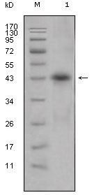
| Calculated MW | 44kDa |
| Description | APOL1(apolipoprotein L, 1), also known as APOL, APO-L, APOL-I. Entrez Protein NP_001130012. It is a 395aa secreted high density lipoprotein which binds to apolipoprotein A-I. And is involved in the formation of most cholesteryl esters in plasma and also promotes efflux of cholesterol from cells. The apolipoprotein L gene family encodes six highly homologous proteins designated apoL-I to -VI. This apolipoprotein L family member may play a role in lipid exchange and transport throughout the body, as well as in reverse cholesterol transport from peripheral cells to the liver. Several different transcript variants encoding different isoforms have been found for this gene. |
| Immunogen | Purified recombinant fragment of APOL1 expressed in E. Coli. |
| Formulation | Ascitic fluid containing 0.03% sodium azide. |
| Gene ID | 8542 |
|---|---|
| Other Names | Apolipoprotein L1, Apolipoprotein L, Apo-L, ApoL, Apolipoprotein L-I, ApoL-I, APOL1, APOL |
| Dilution | WB~~1/500 - 1/2000 E~~N/A |
| Storage | Maintain refrigerated at 2-8°C for up to 6 months. For long term storage store at -20°C in small aliquots to prevent freeze-thaw cycles. |
| Precautions | APOL1 Antibody is for research use only and not for use in diagnostic or therapeutic procedures. |
| Name | APOL1 |
|---|---|
| Synonyms | APOL |
| Function | May play a role in lipid exchange and transport throughout the body. May participate in reverse cholesterol transport from peripheral cells to the liver. |
| Cellular Location | Secreted. |
| Tissue Location | Plasma. Found on APOA-I-containing high density lipoprotein (HDL3). Expressed in pancreas, lung, prostate, liver, placenta and spleen |

Thousands of laboratories across the world have published research that depended on the performance of antibodies from Abcepta to advance their research. Check out links to articles that cite our products in major peer-reviewed journals, organized by research category.
info@abcepta.com, and receive a free "I Love Antibodies" mug.

Provided below are standard protocols that you may find useful for product applications.
References
1. Proc Natl Acad Sci U S A. 2007 Mar 6;104(10):4118-23. 2. J Lipid Res. 2005 Mar;46(3):469-74.
If you have used an Abcepta product and would like to share how it has performed, please click on the "Submit Review" button and provide the requested information. Our staff will examine and post your review and contact you if needed.
If you have any additional inquiries please email technical services at tech@abcepta.com.
Ordering Information
Other Products
Shipping Information
