


 Foundational characteristics of cancer include proliferation, angiogenesis, migration, evasion of apoptosis, and cellular immortality. Find key markers for these cellular processes and antibodies to detect them.
 The SUMOplot™ Analysis Program predicts and scores sumoylation sites in your protein. SUMOylation is a post-translational modification involved in various cellular processes, such as nuclear-cytosolic transport, transcriptional regulation, apoptosis, protein stability, response to stress, and progression through the cell cycle.
 The Autophagy Receptor Motif Plotter predicts and scores autophagy receptor binding sites in your protein. Identifying proteins connected to this pathway is critical to understanding the role of autophagy in physiological as well as pathological processes such as development, differentiation, neurodegenerative diseases, stress, infection, and cancer.
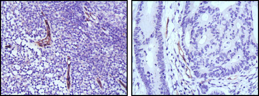
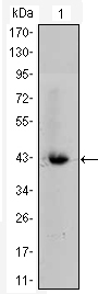
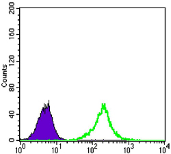
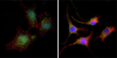
eNOS Antibody
Purified Mouse Monoclonal Antibody

- SPECIFICATION
- CITATIONS
- PROTOCOLS
- BACKGROUND

Application
| IHC, E |
|---|---|
| Primary Accession | P29474 |
| Reactivity | Human |
| Host | Mouse |
| Clonality | Monoclonal |
| Clone Names | 6H2 |
| Isotype | IgG1 |
| Calculated MW | 133kDa |
| Description | Endothelial nitric-oxide synthase (eNOS), also known as NOS3, it is an important enzyme in the cardiovascular system. It is a reactive free radical which acts as a biologic mediator in several processes, including neurotransmission and antimicrobial and antitumoral activities. Nitric oxide is synthesized from L-arginine by nitric oxide synthases. Variations in this gene are associated with susceptibility to coronary spasm. |
| Immunogen | Purified recombinant fragment of human eNOS expressed in E. Coli. |
| Formulation | Ascitic fluid containing 0.03% sodium azide. |
| Gene ID | 4846 |
|---|---|
| Other Names | Nitric oxide synthase, endothelial, 1.14.13.39, Constitutive NOS, cNOS, EC-NOS, Endothelial NOS, eNOS, NOS type III, NOSIII, NOS3 |
| Dilution | IHC~~1/200 - 1/1000 E~~N/A |
| Storage | Maintain refrigerated at 2-8°C for up to 6 months. For long term storage store at -20°C in small aliquots to prevent freeze-thaw cycles. |
| Precautions | eNOS Antibody is for research use only and not for use in diagnostic or therapeutic procedures. |
| Name | NOS3 (HGNC:7876) |
|---|---|
| Function | Produces nitric oxide (NO) which is implicated in vascular smooth muscle relaxation through a cGMP-mediated signal transduction pathway (PubMed:1378832). NO mediates vascular endothelial growth factor (VEGF)-induced angiogenesis in coronary vessels and promotes blood clotting through the activation of platelets. |
| Cellular Location | Cell membrane. Membrane, caveola. Cytoplasm, cytoskeleton. Golgi apparatus. Note=Specifically associates with actin cytoskeleton in the G2 phase of the cell cycle; which is favored by interaction with NOSIP and results in a reduced enzymatic activity |
| Tissue Location | Platelets, placenta, liver and kidney. |

Thousands of laboratories across the world have published research that depended on the performance of antibodies from Abcepta to advance their research. Check out links to articles that cite our products in major peer-reviewed journals, organized by research category.
info@abcepta.com, and receive a free "I Love Antibodies" mug.

Provided below are standard protocols that you may find useful for product applications.
References
1. Nature. 1999 Jun 10;399(6736):601-5. 2. Oncol Rep. 2004 Nov;12(5):1007-11. 3. Breast Cancer Res Treat. 2008 May;109(1):181-2.
If you have used an Abcepta product and would like to share how it has performed, please click on the "Submit Review" button and provide the requested information. Our staff will examine and post your review and contact you if needed.
If you have any additional inquiries please email technical services at tech@abcepta.com.
Ordering Information
Other Products
Shipping Information
