


 Foundational characteristics of cancer include proliferation, angiogenesis, migration, evasion of apoptosis, and cellular immortality. Find key markers for these cellular processes and antibodies to detect them.
 The SUMOplot™ Analysis Program predicts and scores sumoylation sites in your protein. SUMOylation is a post-translational modification involved in various cellular processes, such as nuclear-cytosolic transport, transcriptional regulation, apoptosis, protein stability, response to stress, and progression through the cell cycle.
 The Autophagy Receptor Motif Plotter predicts and scores autophagy receptor binding sites in your protein. Identifying proteins connected to this pathway is critical to understanding the role of autophagy in physiological as well as pathological processes such as development, differentiation, neurodegenerative diseases, stress, infection, and cancer.
RBP4 Antibody
Purified Mouse Monoclonal Antibody
- SPECIFICATION
- CITATIONS
- PROTOCOLS
- BACKGROUND

Application
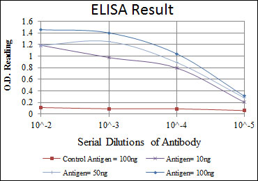
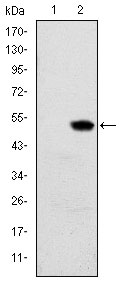
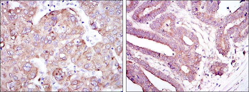
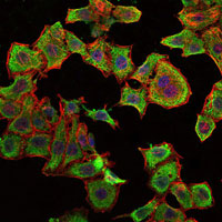
| WB, IHC, FC, ICC, E |
|---|---|
| Primary Accession | P02753 |
| Reactivity | Human |
| Host | Mouse |
| Clonality | Monoclonal |
| Clone Names | 4C2 |
| Isotype | IgG1 |
| Calculated MW | 23kDa |
| Description | This protein belongs to the lipocalin family and is the specific carrier for retinol (vitamin A alcohol) in the blood. It delivers retinol from the liver stores to the peripheral tissues. In plasma, the RBP-retinol complex interacts with transthyretin which prevents its loss by filtration through the kidney glomeruli. A deficiency of vitamin A blocks secretion of the binding protein posttranslationally and results in defective delivery and supply to the epidermal cells. (provided by RefSeq) |
| Immunogen | Purified recombinant fragment of human RBP expressed in E. Coli. |
| Formulation | Ascitic fluid containing 0.03% sodium azide. |
| Gene ID | 5950 |
|---|---|
| Other Names | Retinol-binding protein 4, Plasma retinol-binding protein, PRBP, RBP, Plasma retinol-binding protein(1-182), Plasma retinol-binding protein(1-181), Plasma retinol-binding protein(1-179), Plasma retinol-binding protein(1-176), RBP4 |
| Dilution | WB~~1/500 - 1/2000 IHC~~1/500 - 1/2000 FC~~1/200 - 1/400 ICC~~N/A E~~1/10000 |
| Storage | Maintain refrigerated at 2-8°C for up to 6 months. For long term storage store at -20°C in small aliquots to prevent freeze-thaw cycles. |
| Precautions | RBP4 Antibody is for research use only and not for use in diagnostic or therapeutic procedures. |
| Name | RBP4 |
|---|---|
| Function | Retinol-binding protein that mediates retinol transport in blood plasma (PubMed:5541771). Delivers retinol from the liver stores to the peripheral tissues (Probable). Transfers the bound all-trans retinol to STRA6, that then facilitates retinol transport across the cell membrane (PubMed:22665496). |
| Cellular Location | Secreted |
| Tissue Location | Detected in blood plasma and in urine (at protein level). |

Thousands of laboratories across the world have published research that depended on the performance of antibodies from Abcepta to advance their research. Check out links to articles that cite our products in major peer-reviewed journals, organized by research category.
info@abcepta.com, and receive a free "I Love Antibodies" mug.

Provided below are standard protocols that you may find useful for product applications.
References
1. Diabetologia. 2008 Aug;51(8):1423-8. 2. J Clin Endocrinol Metab. 2008 Aug;93(8):3142-8.
If you have used an Abcepta product and would like to share how it has performed, please click on the "Submit Review" button and provide the requested information. Our staff will examine and post your review and contact you if needed.
If you have any additional inquiries please email technical services at tech@abcepta.com.
Ordering Information
Other Products
Shipping Information
