


 Foundational characteristics of cancer include proliferation, angiogenesis, migration, evasion of apoptosis, and cellular immortality. Find key markers for these cellular processes and antibodies to detect them.
 The SUMOplot™ Analysis Program predicts and scores sumoylation sites in your protein. SUMOylation is a post-translational modification involved in various cellular processes, such as nuclear-cytosolic transport, transcriptional regulation, apoptosis, protein stability, response to stress, and progression through the cell cycle.
 The Autophagy Receptor Motif Plotter predicts and scores autophagy receptor binding sites in your protein. Identifying proteins connected to this pathway is critical to understanding the role of autophagy in physiological as well as pathological processes such as development, differentiation, neurodegenerative diseases, stress, infection, and cancer.
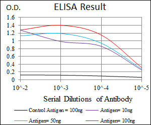
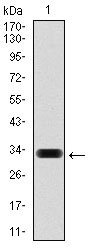
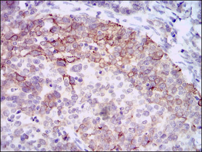
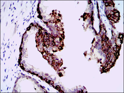
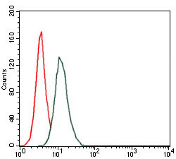
HAS3 Antibody
Purified Mouse Monoclonal Antibody
- SPECIFICATION
- CITATIONS
- PROTOCOLS
- BACKGROUND

Application
| WB, IHC, FC, E |
|---|---|
| Primary Accession | O00219 |
| Reactivity | Human |
| Host | Mouse |
| Clonality | Monoclonal |
| Clone Names | 3C9 |
| Isotype | IgG1 |
| Calculated MW | 63kDa |
| Description | The protein encoded by this gene is involved in the synthesis of the unbranched glycosaminoglycan hyaluronan, or hyaluronic acid, which is a major constituent of the extracellular matrix. This gene is a member of the NODC/HAS gene family. Compared to the proteins encoded by other members of this gene family, this protein appears to be more of a regulator of hyaluronan synthesis. Alternative splicing results in multiple transcript variants. |
| Immunogen | Purified recombinant fragment of human HAS3 expressed in E. Coli. |
| Formulation | Purified antibody in PBS with 0.05% sodium azide |
| Gene ID | 3038 |
|---|---|
| Other Names | Hyaluronan synthase 3, 2.4.1.212, Hyaluronate synthase 3, Hyaluronic acid synthase 3, HA synthase 3, HAS3 |
| Dilution | WB~~1/500 - 1/2000 IHC~~1/200 - 1/1000 FC~~1/200 - 1/400 E~~1/10000 |
| Storage | Maintain refrigerated at 2-8°C for up to 6 months. For long term storage store at -20°C in small aliquots to prevent freeze-thaw cycles. |
| Precautions | HAS3 Antibody is for research use only and not for use in diagnostic or therapeutic procedures. |
| Name | HAS3 (HGNC:4820) |
|---|---|
| Function | Catalyzes the addition of GlcNAc or GlcUA monosaccharides to the nascent hyaluronan polymer. Therefore, it is essential to hyaluronan synthesis a major component of most extracellular matrices that has a structural role in tissues architectures and regulates cell adhesion, migration and differentiation. This is one of three isoenzymes responsible for cellular hyaluronan synthesis. |
| Cellular Location | Cell membrane; Multi-pass membrane protein. Golgi apparatus membrane; Multi-pass membrane protein. Golgi apparatus, trans-Golgi network membrane {ECO:0000250|UniProtKB:O08650}; Multi-pass membrane protein. Early endosome. Note=Travels from endoplasmic reticulum (ER), Golgi to plasma membrane (PubMed:26883802). Actives only when present in plasma membrane (By similarity). O-GlcNAcylation controls its membrane localization (PubMed:26883802). A rapid recycling of HAS3 between plasma membrane and endosomes is controlled by the cytosolic levels of UDP-GlcUA and UDP-GlcNAc (PubMed:26883802) {ECO:0000250|UniProtKB:O08650, ECO:0000269|PubMed:26883802} |
Research Areas
Citations (0)

Thousands of laboratories across the world have published research that depended on the performance of antibodies from Abcepta to advance their research. Check out links to articles that cite our products in major peer-reviewed journals, organized by research category.
Submit your citation using an Abcepta antibody to
info@abcepta.com, and receive a free "I Love Antibodies" mug.
info@abcepta.com, and receive a free "I Love Antibodies" mug.

Application Protocols
Provided below are standard protocols that you may find useful for product applications.
References
BMC Cancer. 2009 May 12;9:143. BMC Cancer. 2010 Sep 27;10:512.
Abcepta welcomes feedback from its customers.
If you have used an Abcepta product and would like to share how it has performed, please click on the "Submit Review" button and provide the requested information. Our staff will examine and post your review and contact you if needed.
If you have any additional inquiries please email technical services at tech@abcepta.com.
$ 435.00
Cat# AO1707a
Ordering Information
United States
AlbaniaAustraliaAustriaBelgiumBosnia & HerzegovinaBrazilBulgariaCanadaCentral AmericaChinaCroatiaCyprusCzech RepublicDenmarkEstoniaFinlandFranceGermanyGreeceHong KongHungaryIcelandIndiaIndonesiaIrelandIsraelItalyJapanLatviaLithuaniaLuxembourgMacedoniaMalaysiaMaltaMexicoNetherlandsNew ZealandNorwayPakistanPolandPortugalRomaniaSerbiaSingaporeSlovakiaSloveniaSouth AfricaSouth KoreaSpainSwedenSwitzerlandTaiwanTurkeyUnited KingdomUnited StatesVietnamWorldwideOthers
USA Headquarters
(888) 735-7227 / (858) 622-0099 or (858) 875-1900
Other Products
Shipping Information
Domestic orders (in stock items)
Shipped out the same day. Orders placed after 1 PM (PST) will ship out the next business day.
International orders
Contact your local distributors
