


 Foundational characteristics of cancer include proliferation, angiogenesis, migration, evasion of apoptosis, and cellular immortality. Find key markers for these cellular processes and antibodies to detect them.
 The SUMOplot™ Analysis Program predicts and scores sumoylation sites in your protein. SUMOylation is a post-translational modification involved in various cellular processes, such as nuclear-cytosolic transport, transcriptional regulation, apoptosis, protein stability, response to stress, and progression through the cell cycle.
 The Autophagy Receptor Motif Plotter predicts and scores autophagy receptor binding sites in your protein. Identifying proteins connected to this pathway is critical to understanding the role of autophagy in physiological as well as pathological processes such as development, differentiation, neurodegenerative diseases, stress, infection, and cancer.
DLL4 Antibody
Purified Mouse Monoclonal Antibody
- SPECIFICATION
- CITATIONS
- PROTOCOLS
- BACKGROUND

Application
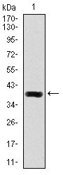
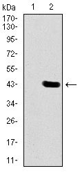
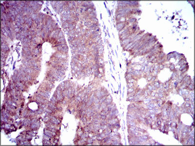
| WB, IHC, E |
|---|---|
| Primary Accession | Q9NR61 |
| Reactivity | Human |
| Host | Mouse |
| Clonality | Monoclonal |
| Clone Names | 4A11F8 |
| Isotype | IgG1 |
| Calculated MW | 74.6kDa |
| Description | This gene is a homolog of the Drosophila delta gene. The delta gene family encodes Notch ligands that are characterized by a DSL domain, EGF repeats, and a transmembrane domain. |
| Immunogen | Purified recombinant fragment of human DLL4 (AA: 313-439) expressed in E. Coli. |
| Formulation | Purified antibody in PBS with 0.05% sodium azide |
| Gene ID | 54567 |
|---|---|
| Other Names | Delta-like protein 4, Drosophila Delta homolog 4, Delta4, DLL4 |
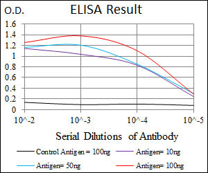
| Dilution | WB~~1/500 - 1/2000 IHC~~1/200 - 1/1000 E~~1/10000 |
| Storage | Maintain refrigerated at 2-8°C for up to 6 months. For long term storage store at -20°C in small aliquots to prevent freeze-thaw cycles. |
| Precautions | DLL4 Antibody is for research use only and not for use in diagnostic or therapeutic procedures. |
| Name | DLL4 |
|---|---|
| Function | Involved in the Notch signaling pathway as Notch ligand (PubMed:11134954). Activates NOTCH1 and NOTCH4. Involved in angiogenesis; negatively regulates endothelial cell proliferation and migration and angiogenic sprouting (PubMed:20616313). Essential for retinal progenitor proliferation. Required for suppressing rod fates in late retinal progenitors as well as for proper generation of other retinal cell types (By similarity). During spinal cord neurogenesis, inhibits V2a interneuron fate (PubMed:17728344). |
| Cellular Location | Cell membrane; Single-pass type I membrane protein |
| Tissue Location | Expressed in vascular endothelium. |

Thousands of laboratories across the world have published research that depended on the performance of antibodies from Abcepta to advance their research. Check out links to articles that cite our products in major peer-reviewed journals, organized by research category.
info@abcepta.com, and receive a free "I Love Antibodies" mug.

Provided below are standard protocols that you may find useful for product applications.
References
1.Am J Pathol. 2010 Apr;176(4):2019-28. 2.Blood. 2010 Sep 30;116(13):2385-94.
If you have used an Abcepta product and would like to share how it has performed, please click on the "Submit Review" button and provide the requested information. Our staff will examine and post your review and contact you if needed.
If you have any additional inquiries please email technical services at tech@abcepta.com.
Ordering Information
Other Products
Shipping Information
