


 Foundational characteristics of cancer include proliferation, angiogenesis, migration, evasion of apoptosis, and cellular immortality. Find key markers for these cellular processes and antibodies to detect them.
 The SUMOplot™ Analysis Program predicts and scores sumoylation sites in your protein. SUMOylation is a post-translational modification involved in various cellular processes, such as nuclear-cytosolic transport, transcriptional regulation, apoptosis, protein stability, response to stress, and progression through the cell cycle.
 The Autophagy Receptor Motif Plotter predicts and scores autophagy receptor binding sites in your protein. Identifying proteins connected to this pathway is critical to understanding the role of autophagy in physiological as well as pathological processes such as development, differentiation, neurodegenerative diseases, stress, infection, and cancer.
CFLAR Antibody
Purified Mouse Monoclonal Antibody
- SPECIFICATION
- CITATIONS
- PROTOCOLS
- BACKGROUND

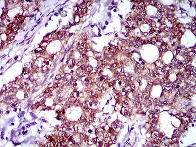
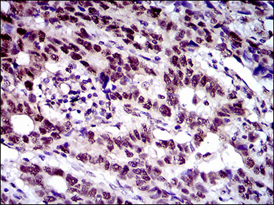
Application
| WB, IHC, E |
|---|---|
| Primary Accession | O15519 |
| Reactivity | Human |
| Host | Mouse |
| Clonality | Monoclonal |
| Clone Names | 6G11A6 |
| Isotype | IgG1 |
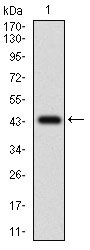
| Calculated MW | 55.3kDa |
| Description | The protein encoded by this gene is a regulator of apoptosis and is structurally similar to caspase-8. However, the encoded protein lacks caspase activity and appears to be itself cleaved into two peptides by caspase-8. Several transcript variants encoding different isoforms have been found for this gene, and partial evidence for several more variants exists. |
| Immunogen | Purified recombinant fragment of human CFLAR (AA: 100-251) expressed in E. Coli. |
| Formulation | Purified antibody in PBS with 0.05% sodium azide |
| Gene ID | 8837 |
|---|---|
| Other Names | CASP8 and FADD-like apoptosis regulator, Caspase homolog, CASH, Caspase-eight-related protein, Casper, Caspase-like apoptosis regulatory protein, CLARP, Cellular FLICE-like inhibitory protein, c-FLIP, FADD-like antiapoptotic molecule 1, FLAME-1, Inhibitor of FLICE, I-FLICE, MACH-related inducer of toxicity, MRIT, Usurpin, CASP8 and FADD-like apoptosis regulator subunit p43, CASP8 and FADD-like apoptosis regulator subunit p12, CFLAR, CASH, CASP8AP1, CLARP, MRIT |
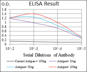
| Dilution | WB~~1/500 - 1/2000 IHC~~1/200 - 1/1000 E~~1/10000 |
| Storage | Maintain refrigerated at 2-8°C for up to 6 months. For long term storage store at -20°C in small aliquots to prevent freeze-thaw cycles. |
| Precautions | CFLAR Antibody is for research use only and not for use in diagnostic or therapeutic procedures. |
| Name | CFLAR |
|---|---|
| Synonyms | CASH, CASP8AP1, CLARP, MRIT |
| Function | Apoptosis regulator protein which may function as a crucial link between cell survival and cell death pathways in mammalian cells. Acts as an inhibitor of TNFRSF6 mediated apoptosis. A proteolytic fragment (p43) is likely retained in the death-inducing signaling complex (DISC) thereby blocking further recruitment and processing of caspase-8 at the complex. Full length and shorter isoforms have been shown either to induce apoptosis or to reduce TNFRSF-triggered apoptosis. Lacks enzymatic (caspase) activity. |
| Tissue Location | Widely expressed. Higher expression in skeletal muscle, pancreas, heart, kidney, placenta, and peripheral blood leukocytes. Also detected in diverse cell lines. Isoform 8 is predominantly expressed in testis and skeletal muscle |

Thousands of laboratories across the world have published research that depended on the performance of antibodies from Abcepta to advance their research. Check out links to articles that cite our products in major peer-reviewed journals, organized by research category.
info@abcepta.com, and receive a free "I Love Antibodies" mug.

Provided below are standard protocols that you may find useful for product applications.
Background
The protein encoded by this gene is a regulator of apoptosis and is structurally similar to caspase-8. However, the encoded protein lacks caspase activity and appears to be itself cleaved into two peptides by caspase-8. Several transcript variants encoding different isoforms have been found for this gene, and partial evidence for several more variants exists. ; ;
References
1. Pediatr Blood Cancer. 2013 Apr;60(4):575-9. 2. J Cell Biochem. 2012 Dec;113(12):3692-700.
If you have used an Abcepta product and would like to share how it has performed, please click on the "Submit Review" button and provide the requested information. Our staff will examine and post your review and contact you if needed.
If you have any additional inquiries please email technical services at tech@abcepta.com.
Ordering Information
Other Products
Shipping Information
