


 Foundational characteristics of cancer include proliferation, angiogenesis, migration, evasion of apoptosis, and cellular immortality. Find key markers for these cellular processes and antibodies to detect them.
 The SUMOplot™ Analysis Program predicts and scores sumoylation sites in your protein. SUMOylation is a post-translational modification involved in various cellular processes, such as nuclear-cytosolic transport, transcriptional regulation, apoptosis, protein stability, response to stress, and progression through the cell cycle.
 The Autophagy Receptor Motif Plotter predicts and scores autophagy receptor binding sites in your protein. Identifying proteins connected to this pathway is critical to understanding the role of autophagy in physiological as well as pathological processes such as development, differentiation, neurodegenerative diseases, stress, infection, and cancer.
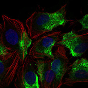
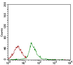
CD74 Antibody
Purified Mouse Monoclonal Antibody
- SPECIFICATION
- CITATIONS
- PROTOCOLS
- BACKGROUND

Application
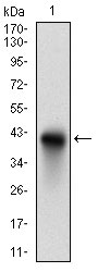
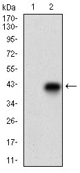
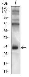
| WB, FC, ICC, E |
|---|---|
| Primary Accession | P04233 |
| Reactivity | Human |
| Host | Mouse |
| Clonality | Monoclonal |
| Clone Names | 2D1B3 |
| Isotype | IgG1 |
| Calculated MW | 33.5kDa |
| Description | The protein encoded by this gene associates with class II major histocompatibility complex (MHC) and is an important chaperone that regulates antigen presentation for immune response. It also serves as cell surface receptor for the cytokine macrophage migration inhibitory factor (MIF) which, when bound to the encoded protein, initiates survival pathways and cell proliferation. This protein also interacts with amyloid precursor protein (APP) and suppresses the production of amyloid beta (Abeta). Multiple alternatively spliced transcript variants encoding different isoforms have been identified. |
| Immunogen | Purified recombinant fragment of human CD74 (AA: 1-106) expressed in E. Coli. |
| Formulation | Purified antibody in PBS with 0.05% sodium azide |
| Gene ID | 972 |
|---|---|
| Other Names | HLA class II histocompatibility antigen gamma chain, HLA-DR antigens-associated invariant chain, Ia antigen-associated invariant chain, Ii, p33, CD74, CD74, DHLAG |
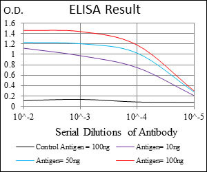
| Dilution | WB~~1/500 - 1/2000 FC~~1/200 - 1/400 ICC~~N/A E~~1/10000 |
| Storage | Maintain refrigerated at 2-8°C for up to 6 months. For long term storage store at -20°C in small aliquots to prevent freeze-thaw cycles. |
| Precautions | CD74 Antibody is for research use only and not for use in diagnostic or therapeutic procedures. |
| Name | CD74 (HGNC:1697) |
|---|---|
| Synonyms | DHLAG |
| Function | Plays a critical role in MHC class II antigen processing by stabilizing peptide-free class II alpha/beta heterodimers in a complex soon after their synthesis and directing transport of the complex from the endoplasmic reticulum to the endosomal/lysosomal system where the antigen processing and binding of antigenic peptides to MHC class II takes place. Serves as cell surface receptor for the cytokine MIF. [Isoform p41]: Stabilizes the conformation of mature CTSL by binding to its active site and serving as a chaperone to help maintain a pool of mature enzyme in endocytic compartments and extracellular space of antigen-presenting cells (APCs). Has antiviral activity by stymieing the endosomal entry of Ebola virus and coronaviruses, including SARS-CoV-2 (PubMed:32855215). Disrupts cathepsin-mediated Ebola virus glycoprotein processing, which prevents viral fusion and entry. This antiviral activity is specific to p41 isoform (PubMed:32855215). |
| Cellular Location | Cell membrane; Single-pass type II membrane protein. Endoplasmic reticulum membrane. Golgi apparatus, trans-Golgi network. Endosome. Lysosome. Secreted. Note=Transits through a number of intracellular compartments in the endocytic pathway. It can either undergo proteolysis or reach the cell membrane |
| Tissue Location | Detected in urine (at protein level). [Isoform p33]: In B cells, represents 70% of total CD74 expression. |

Thousands of laboratories across the world have published research that depended on the performance of antibodies from Abcepta to advance their research. Check out links to articles that cite our products in major peer-reviewed journals, organized by research category.
info@abcepta.com, and receive a free "I Love Antibodies" mug.

Provided below are standard protocols that you may find useful for product applications.
Background
The protein encoded by this gene is a transcriptional activator. It can bind the consensus DNA sequence 5'-DHWATTGAYTWWD-3' on a variety of gene promoters such as those of HNF3B and TTR(By similarity). ;
References
1. Tumour Biol. 2012 Dec;33(6):2273-7. 2. World J Gastroenterol. 2012 May 14;18(18):2253-61.
If you have used an Abcepta product and would like to share how it has performed, please click on the "Submit Review" button and provide the requested information. Our staff will examine and post your review and contact you if needed.
If you have any additional inquiries please email technical services at tech@abcepta.com.
Ordering Information
Other Products
Shipping Information
