


 Foundational characteristics of cancer include proliferation, angiogenesis, migration, evasion of apoptosis, and cellular immortality. Find key markers for these cellular processes and antibodies to detect them.
 The SUMOplot™ Analysis Program predicts and scores sumoylation sites in your protein. SUMOylation is a post-translational modification involved in various cellular processes, such as nuclear-cytosolic transport, transcriptional regulation, apoptosis, protein stability, response to stress, and progression through the cell cycle.
 The Autophagy Receptor Motif Plotter predicts and scores autophagy receptor binding sites in your protein. Identifying proteins connected to this pathway is critical to understanding the role of autophagy in physiological as well as pathological processes such as development, differentiation, neurodegenerative diseases, stress, infection, and cancer.
PLA2G12A Antibody
Purified Mouse Monoclonal Antibody
- SPECIFICATION
- CITATIONS
- PROTOCOLS
- BACKGROUND

Application
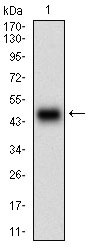
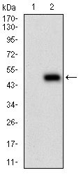
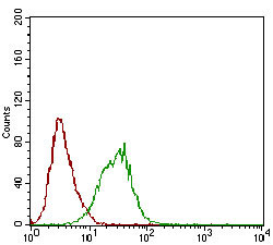
| WB, FC, E |
|---|---|
| Primary Accession | Q9BZM1 |
| Reactivity | Human |
| Host | Mouse |
| Clonality | Monoclonal |
| Clone Names | 7C7C9 |
| Isotype | IgG1 |
| Calculated MW | 21kDa |
| Description | Secreted phospholipase A2 (sPLA2) enzymes liberate arachidonic acid from phospholipids for production of eicosanoids and exert a variety of physiologic and pathologic effects. Group XII sPLA2s, such as PLA2G12A, have relatively low specific activity and are structurally and functionally distinct from other sPLA2s |
| Immunogen | Purified recombinant fragment of human PLA2G12A (AA: 21-189) expressed in E. Coli. |
| Formulation | Purified antibody in PBS with 0.05% sodium azide. |
| Gene ID | 81579 |
|---|---|
| Other Names | Group XIIA secretory phospholipase A2, GXII sPLA2, sPLA2-XII, 3.1.1.4, Phosphatidylcholine 2-acylhydrolase 12A, PLA2G12A, PLA2G12 |
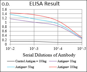
| Dilution | WB~~1/500 - 1/2000 FC~~1/200 - 1/400 E~~1/10000 |
| Storage | Maintain refrigerated at 2-8°C for up to 6 months. For long term storage store at -20°C in small aliquots to prevent freeze-thaw cycles. |
| Precautions | PLA2G12A Antibody is for research use only and not for use in diagnostic or therapeutic procedures. |
| Name | PLA2G12A |
|---|---|
| Synonyms | PLA2G12 |
| Function | PA2 catalyzes the calcium-dependent hydrolysis of the 2-acyl groups in 3-sn-phosphoglycerides. Does not exhibit detectable activity toward sn-2-arachidonoyl- or linoleoyl-phosphatidylcholine or -phosphatidylethanolamine. |
| Cellular Location | Secreted. Cytoplasm |
| Tissue Location | Abundantly expressed in heart, skeletal muscle, kidney, liver and pancreas |

Thousands of laboratories across the world have published research that depended on the performance of antibodies from Abcepta to advance their research. Check out links to articles that cite our products in major peer-reviewed journals, organized by research category.
info@abcepta.com, and receive a free "I Love Antibodies" mug.

Provided below are standard protocols that you may find useful for product applications.
Background
The protein encoded by this gene is found in the nucleus and is a cofactor of DNA polymerase delta. The encoded protein acts as a homotrimer and helps increase the processivity of leading strand synthesis during DNA replication. In response to DNA damage, this protein is ubiquitinated and is involved in the RAD6-dependent DNA repair pathway. Two transcript variants encoding the same protein have been found for this gene. Pseudogenes of this gene have been described on chromosome 4 and on the X chromosome. ; ;
References
1. J Biol Chem. 2003 Mar 21;278(12):10657-67. 2. Biochemistry. 2003 Oct 7;42(39):11494-503.
If you have used an Abcepta product and would like to share how it has performed, please click on the "Submit Review" button and provide the requested information. Our staff will examine and post your review and contact you if needed.
If you have any additional inquiries please email technical services at tech@abcepta.com.
Ordering Information
Other Products
Shipping Information
