


 Foundational characteristics of cancer include proliferation, angiogenesis, migration, evasion of apoptosis, and cellular immortality. Find key markers for these cellular processes and antibodies to detect them.
 The SUMOplot™ Analysis Program predicts and scores sumoylation sites in your protein. SUMOylation is a post-translational modification involved in various cellular processes, such as nuclear-cytosolic transport, transcriptional regulation, apoptosis, protein stability, response to stress, and progression through the cell cycle.
 The Autophagy Receptor Motif Plotter predicts and scores autophagy receptor binding sites in your protein. Identifying proteins connected to this pathway is critical to understanding the role of autophagy in physiological as well as pathological processes such as development, differentiation, neurodegenerative diseases, stress, infection, and cancer.
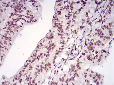
MEN1 Antibody
Purified Mouse Monoclonal Antibody
- SPECIFICATION
- CITATIONS
- PROTOCOLS
- BACKGROUND

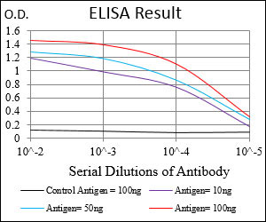
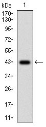
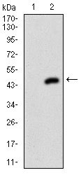
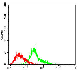
Application
| WB, IHC, FC, E |
|---|---|
| Primary Accession | O00255 |
| Reactivity | Human |
| Host | Mouse |
| Clonality | Monoclonal |
| Clone Names | 7D3E10 |
| Isotype | IgG1 |
| Calculated MW | 68kDa |
| Description | This gene encodes menin, a putative tumor suppressor associated with a syndrome known as multiple endocrine neoplasia type 1. In vitro studies have shown menin is localized to the nucleus, possesses two functional nuclear localization signals, and inhibits transcriptional activation by JunD, however, the function of this protein is not known. Two messages have been detected on northern blots but the larger message has not been characterized. Alternative splicing results in multiple transcript variants. |
| Immunogen | Purified recombinant fragment of human MEN1 (AA: 392-554) expressed in E. Coli. |
| Formulation | Purified antibody in PBS with 0.05% sodium azide. |
| Gene ID | 4221 |
|---|---|
| Other Names | Menin, MEN1, SCG2 |
| Dilution | WB~~1/500 - 1/2000 IHC~~1/200 - 1/1000 FC~~1/200 - 1/400 E~~1/10000 |
| Storage | Maintain refrigerated at 2-8°C for up to 6 months. For long term storage store at -20°C in small aliquots to prevent freeze-thaw cycles. |
| Precautions | MEN1 Antibody is for research use only and not for use in diagnostic or therapeutic procedures. |
| Name | MEN1 |
|---|---|
| Synonyms | SCG2 |
| Function | Essential component of a MLL/SET1 histone methyltransferase (HMT) complex, a complex that specifically methylates 'Lys-4' of histone H3 (H3K4). Functions as a transcriptional regulator. Binds to the TERT promoter and represses telomerase expression. Plays a role in TGFB1-mediated inhibition of cell-proliferation, possibly regulating SMAD3 transcriptional activity. Represses JUND-mediated transcriptional activation on AP1 sites, as well as that mediated by NFKB subunit RELA. Positively regulates HOXC8 and HOXC6 gene expression. May be involved in normal hematopoiesis through the activation of HOXA9 expression (By similarity). May be involved in DNA repair. |
| Cellular Location | Nucleus. Note=Concentrated in nuclear body-like structures. Relocates to the nuclear matrix upon gamma irradiation |
| Tissue Location | Ubiquitous. |

Thousands of laboratories across the world have published research that depended on the performance of antibodies from Abcepta to advance their research. Check out links to articles that cite our products in major peer-reviewed journals, organized by research category.
info@abcepta.com, and receive a free "I Love Antibodies" mug.

Provided below are standard protocols that you may find useful for product applications.
Background
The innate immune system confers host defense against viral and microbial infection, and TRAFD1 is a negative feedback regulator that controls excessive immune responses ; ;
References
1. Clinics (Sao Paulo). 2012;67 Suppl 1:49-56.2. World J Surg Oncol. 2011 Jan 25;9:6.
If you have used an Abcepta product and would like to share how it has performed, please click on the "Submit Review" button and provide the requested information. Our staff will examine and post your review and contact you if needed.
If you have any additional inquiries please email technical services at tech@abcepta.com.
Ordering Information
Other Products
Shipping Information
