


 Foundational characteristics of cancer include proliferation, angiogenesis, migration, evasion of apoptosis, and cellular immortality. Find key markers for these cellular processes and antibodies to detect them.
 The SUMOplot™ Analysis Program predicts and scores sumoylation sites in your protein. SUMOylation is a post-translational modification involved in various cellular processes, such as nuclear-cytosolic transport, transcriptional regulation, apoptosis, protein stability, response to stress, and progression through the cell cycle.
 The Autophagy Receptor Motif Plotter predicts and scores autophagy receptor binding sites in your protein. Identifying proteins connected to this pathway is critical to understanding the role of autophagy in physiological as well as pathological processes such as development, differentiation, neurodegenerative diseases, stress, infection, and cancer.
FTL Antibody
Purified Mouse Monoclonal Antibody

- SPECIFICATION
- CITATIONS
- PROTOCOLS
- BACKGROUND

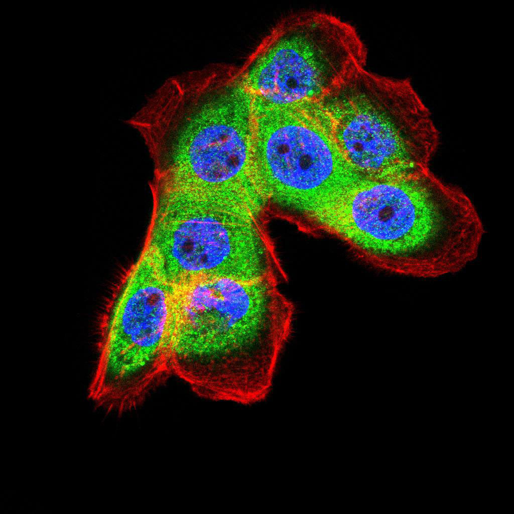
Application
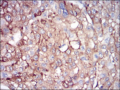
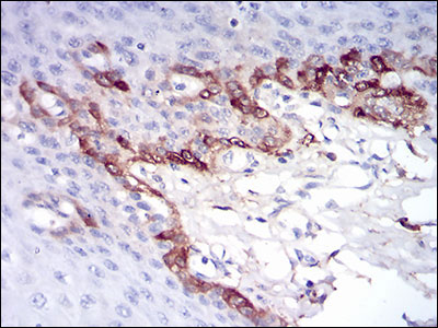
| WB, IHC, ICC, E |
|---|---|
| Primary Accession | P02792 |
| Reactivity | Human |
| Host | Mouse |
| Clonality | Monoclonal |
| Clone Names | 8E1E7 |
| Isotype | IgG1 |
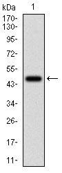
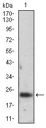
| Calculated MW | 20kDa |
| Description | This gene encodes the light subunit of the ferritin protein. Ferritin is the major intracellular iron storage protein in prokaryotes and eukaryotes. It is composed of 24 subunits of the heavy and light ferritin chains. Variation in ferritin subunit composition may affect the rates of iron uptake and release in different tissues. A major function of ferritin is the storage of iron in a soluble and nontoxic state. Defects in this light chain ferritin gene are associated with several neurodegenerative diseases and hyperferritinemia-cataract syndrome. This gene has multiple pseudogenes. |
| Immunogen | Purified recombinant fragment of human FTL (AA: FULL(1-157)) expressed in E. Coli. |
| Formulation | Purified antibody in PBS with 0.05% sodium azide. |
| Gene ID | 2512 |
|---|---|
| Other Names | Ferritin light chain, Ferritin L subunit, FTL |
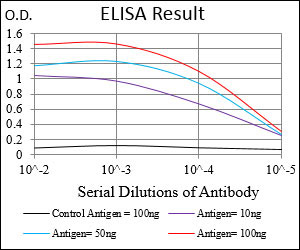
| Dilution | WB~~1/500 - 1/2000 IHC~~1/200 - 1/1000 ICC~~N/A E~~1/10000 |
| Storage | Maintain refrigerated at 2-8°C for up to 6 months. For long term storage store at -20°C in small aliquots to prevent freeze-thaw cycles. |
| Precautions | FTL Antibody is for research use only and not for use in diagnostic or therapeutic procedures. |
| Name | FTL |
|---|---|
| Function | Stores iron in a soluble, non-toxic, readily available form. Important for iron homeostasis. Iron is taken up in the ferrous form and deposited as ferric hydroxides after oxidation. Also plays a role in delivery of iron to cells. Mediates iron uptake in capsule cells of the developing kidney (By similarity). Delivery to lysosomes by the cargo receptor NCOA4 for autophagic degradation and release or iron (PubMed:24695223). |
| Cellular Location | Cytoplasmic vesicle, autophagosome. Cytoplasm {ECO:0000250|UniProtKB:P29391}. Autolysosome {ECO:0000250|UniProtKB:P29391} |

Thousands of laboratories across the world have published research that depended on the performance of antibodies from Abcepta to advance their research. Check out links to articles that cite our products in major peer-reviewed journals, organized by research category.
info@abcepta.com, and receive a free "I Love Antibodies" mug.

Provided below are standard protocols that you may find useful for product applications.
Background
The protein encoded by this gene belongs to the cyclic nucleotide phosphodiesterase (PDE) family, and PDE1 subfamily. Members of the PDE1 family are calmodulin-dependent PDEs that are stimulated by a calcium-calmodulin complex. This PDE has dual-specificity for the second messengers, cAMP and cGMP, with a preference for cGMP as a substrate. cAMP and cGMP function as key regulators of many important physiological processes. Alternatively spliced transcript variants encoding different isoforms have been described for this gene. ; ;
References
1. Free Radic Biol Med. 2012 May 1;52(9):1692-7.2. Neurobiol Dis. 2010 Jan;37(1):77-85.
If you have used an Abcepta product and would like to share how it has performed, please click on the "Submit Review" button and provide the requested information. Our staff will examine and post your review and contact you if needed.
If you have any additional inquiries please email technical services at tech@abcepta.com.
Ordering Information
Other Products
Shipping Information
