


 Foundational characteristics of cancer include proliferation, angiogenesis, migration, evasion of apoptosis, and cellular immortality. Find key markers for these cellular processes and antibodies to detect them.
 The SUMOplot™ Analysis Program predicts and scores sumoylation sites in your protein. SUMOylation is a post-translational modification involved in various cellular processes, such as nuclear-cytosolic transport, transcriptional regulation, apoptosis, protein stability, response to stress, and progression through the cell cycle.
 The Autophagy Receptor Motif Plotter predicts and scores autophagy receptor binding sites in your protein. Identifying proteins connected to this pathway is critical to understanding the role of autophagy in physiological as well as pathological processes such as development, differentiation, neurodegenerative diseases, stress, infection, and cancer.
PTPN1 Antibody
Purified Mouse Monoclonal Antibody
- SPECIFICATION
- CITATIONS
- PROTOCOLS
- BACKGROUND

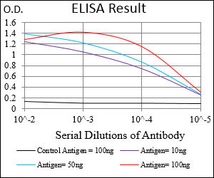
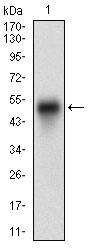
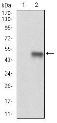
Application
| WB, E |
|---|---|
| Primary Accession | P18031 |
| Reactivity | Human |
| Host | Mouse |
| Clonality | Monoclonal |
| Clone Names | 4F8F2 |
| Isotype | IgG2b |
| Calculated MW | 50kDa |
| Description | The protein encoded by this gene is the founding member of the protein tyrosine phosphatase (PTP) family, which was isolated and identified based on its enzymatic activity and amino acid sequence. PTPs catalyze the hydrolysis of the phosphate monoesters specifically on tyrosine residues. Members of the PTP family share a highly conserved catalytic motif, which is essential for the catalytic activity. PTPs are known to be signaling molecules that regulate a variety of cellular processes including cell growth, differentiation, mitotic cycle, and oncogenic transformation. This PTP has been shown to act as a negative regulator of insulin signaling by dephosphorylating the phosphotryosine residues of insulin receptor kinase. This PTP was also reported to dephosphorylate epidermal growth factor receptor kinase, as well as JAK2 and TYK2 kinases, which implicated the role of this PTP in cell growth control, and cell response to interferon stimulation. Two transcript variants encoding different isoforms have been found for this gene. |
| Immunogen | Purified recombinant fragment of human PTPN1 (AA: 40-246) expressed in E. Coli. |
| Formulation | Purified antibody in PBS with 0.05% sodium azide. |
| Gene ID | 5770 |
|---|---|
| Other Names | Tyrosine-protein phosphatase non-receptor type 1, 3.1.3.48, Protein-tyrosine phosphatase 1B, PTP-1B, PTPN1, PTP1B |
| Dilution | WB~~1/500 - 1/2000 E~~1/10000 |
| Storage | Maintain refrigerated at 2-8°C for up to 6 months. For long term storage store at -20°C in small aliquots to prevent freeze-thaw cycles. |
| Precautions | PTPN1 Antibody is for research use only and not for use in diagnostic or therapeutic procedures. |
| Name | PTPN1 |
|---|---|
| Synonyms | PTP1B |
| Function | Tyrosine-protein phosphatase which acts as a regulator of endoplasmic reticulum unfolded protein response. Mediates dephosphorylation of EIF2AK3/PERK; inactivating the protein kinase activity of EIF2AK3/PERK. May play an important role in CKII- and p60c- src-induced signal transduction cascades. May regulate the EFNA5-EPHA3 signaling pathway which modulates cell reorganization and cell-cell repulsion. May also regulate the hepatocyte growth factor receptor signaling pathway through dephosphorylation of MET. |
| Cellular Location | Endoplasmic reticulum membrane; Peripheral membrane protein; Cytoplasmic side Note=Interacts with EPHA3 at the cell membrane |
| Tissue Location | Expressed in keratinocytes (at protein level). |

Thousands of laboratories across the world have published research that depended on the performance of antibodies from Abcepta to advance their research. Check out links to articles that cite our products in major peer-reviewed journals, organized by research category.
info@abcepta.com, and receive a free "I Love Antibodies" mug.

Provided below are standard protocols that you may find useful for product applications.
Background
This gene is expressed ubiquitously with higher levels in fetal than in adult tissues. It encodes a protein sharing 93% sequence identity with the mouse protein. Wolf-Hirschhorn syndrome (WHS) is a malformation syndrome associated with a hemizygous deletion of the distal short arm of chromosome 4. This gene is mapped to the 165 kb WHS critical region, and may play a role in the phenotype of the WHS or Pitt-Rogers-Danks syndrome. The encoded protein is found to be capable of reacting with HLA-A2-restricted and tumor-specific cytotoxic T lymphocytes, suggesting a target for use in specific immunotherapy for a large number of cancer patients. This protein has also been shown to be a member of the NELF (negative elongation factor) protein complex that participates in the regulation of RNA polymerase II transcription elongation. ; ;
References
1. Med Oncol. 2012 Jun;29(2):948-56. 2. Cell Biol Int. 2010 Jul;34(7):747-53.
If you have used an Abcepta product and would like to share how it has performed, please click on the "Submit Review" button and provide the requested information. Our staff will examine and post your review and contact you if needed.
If you have any additional inquiries please email technical services at tech@abcepta.com.
Ordering Information
Other Products
Shipping Information
