


 Foundational characteristics of cancer include proliferation, angiogenesis, migration, evasion of apoptosis, and cellular immortality. Find key markers for these cellular processes and antibodies to detect them.
 The SUMOplot™ Analysis Program predicts and scores sumoylation sites in your protein. SUMOylation is a post-translational modification involved in various cellular processes, such as nuclear-cytosolic transport, transcriptional regulation, apoptosis, protein stability, response to stress, and progression through the cell cycle.
 The Autophagy Receptor Motif Plotter predicts and scores autophagy receptor binding sites in your protein. Identifying proteins connected to this pathway is critical to understanding the role of autophagy in physiological as well as pathological processes such as development, differentiation, neurodegenerative diseases, stress, infection, and cancer.
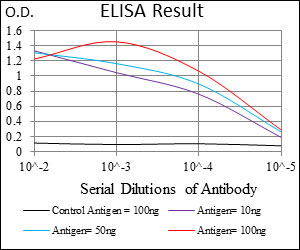
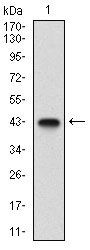
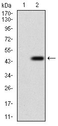
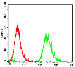
EPN1 Antibody
Purified Mouse Monoclonal Antibody
- SPECIFICATION
- CITATIONS
- PROTOCOLS
- BACKGROUND

Application
| WB, FC, E |
|---|---|
| Primary Accession | Q9Y6I3 |
| Reactivity | Human |
| Host | Mouse |
| Clonality | Monoclonal |
| Clone Names | 6F7F9 |
| Isotype | IgG1 |
| Calculated MW | 60.3kDa |
| Description | The protein encoded by this gene binds clathrin and is involved in the endocytosis of clathrin-coated vesicles. Three transcript variants encoding different isoforms have been found for this gene. |
| Immunogen | Purified recombinant fragment of human EPN1 (AA: 106-254) expressed in E. Coli. |
| Formulation | Purified antibody in PBS with 0.05% sodium azide. |
| Gene ID | 29924 |
|---|---|
| Other Names | Epsin-1, EH domain-binding mitotic phosphoprotein, EPS-15-interacting protein 1, EPN1 |
| Dilution | WB~~1/500 - 1/2000 FC~~1/200 - 1/400 E~~1/10000 |
| Storage | Maintain refrigerated at 2-8°C for up to 6 months. For long term storage store at -20°C in small aliquots to prevent freeze-thaw cycles. |
| Precautions | EPN1 Antibody is for research use only and not for use in diagnostic or therapeutic procedures. |
| Name | EPN1 |
|---|---|
| Function | Binds to membranes enriched in phosphatidylinositol 4,5- bisphosphate (PtdIns(4,5)P2). Modifies membrane curvature and facilitates the formation of clathrin-coated invaginations (By similarity). Regulates receptor-mediated endocytosis (PubMed:10393179, PubMed:10557078). |
| Cellular Location | Cytoplasm. Cell membrane; Peripheral membrane protein. Nucleus. Membrane, clathrin-coated pit Note=Associated with the cytoplasmic membrane at sites where clathrin- coated pits are forming. Colocalizes with clathrin and AP-2 in a punctate pattern on the plasma membrane. Detected in presynaptic nerve terminals and in Golgi stacks. May shuttle to the nucleus when associated with ZBTB16/ZNF145 (By similarity). |

Thousands of laboratories across the world have published research that depended on the performance of antibodies from Abcepta to advance their research. Check out links to articles that cite our products in major peer-reviewed journals, organized by research category.
info@abcepta.com, and receive a free "I Love Antibodies" mug.

Provided below are standard protocols that you may find useful for product applications.
References
1. J Biol Chem. 2011 Nov 25;286(47):40760-70. 2. Traffic. 2009 Feb;10(2):235-45.
If you have used an Abcepta product and would like to share how it has performed, please click on the "Submit Review" button and provide the requested information. Our staff will examine and post your review and contact you if needed.
If you have any additional inquiries please email technical services at tech@abcepta.com.
Ordering Information
Other Products
Shipping Information
