


 Foundational characteristics of cancer include proliferation, angiogenesis, migration, evasion of apoptosis, and cellular immortality. Find key markers for these cellular processes and antibodies to detect them.
 The SUMOplot™ Analysis Program predicts and scores sumoylation sites in your protein. SUMOylation is a post-translational modification involved in various cellular processes, such as nuclear-cytosolic transport, transcriptional regulation, apoptosis, protein stability, response to stress, and progression through the cell cycle.
 The Autophagy Receptor Motif Plotter predicts and scores autophagy receptor binding sites in your protein. Identifying proteins connected to this pathway is critical to understanding the role of autophagy in physiological as well as pathological processes such as development, differentiation, neurodegenerative diseases, stress, infection, and cancer.
PRDM1 Antibody
Purified Mouse Monoclonal Antibody
- SPECIFICATION
- CITATIONS
- PROTOCOLS
- BACKGROUND

Application
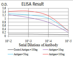
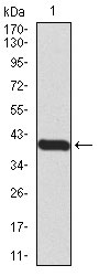
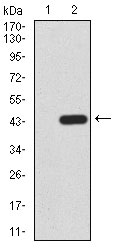
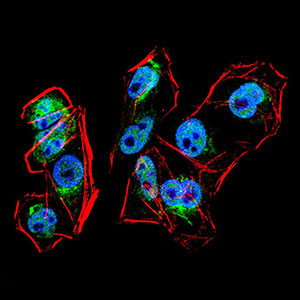
| WB, ICC, E |
|---|---|
| Primary Accession | O75626 |
| Reactivity | Human |
| Host | Mouse |
| Clonality | Monoclonal |
| Clone Names | 7C11H1 |
| Isotype | IgG2a |
| Calculated MW | 91.7kDa |
| Description | This gene encodes a protein that acts as a repressor of beta-interferon gene expression. The protein binds specifically to the PRDI (positive regulatory domain I element) of the beta-IFN gene promoter. Transcription of this gene increases upon virus induction. Two alternatively spliced transcript variants that encode different isoforms have been reported. |
| Immunogen | Purified recombinant fragment of human PRDM1 (AA: 690-825) expressed in E. Coli. |
| Formulation | Purified antibody in PBS with 0.05% sodium azide |
| Gene ID | 639 |
|---|---|
| Other Names | PR domain zinc finger protein 1, 2.1.1.-, BLIMP-1, Beta-interferon gene positive regulatory domain I-binding factor, PR domain-containing protein 1, Positive regulatory domain I-binding factor 1, PRDI-BF1, PRDI-binding factor 1, PRDM1, BLIMP1 |
| Dilution | WB~~1/500 - 1/2000 ICC~~N/A E~~1/10000 |
| Storage | Maintain refrigerated at 2-8°C for up to 6 months. For long term storage store at -20°C in small aliquots to prevent freeze-thaw cycles. |
| Precautions | PRDM1 Antibody is for research use only and not for use in diagnostic or therapeutic procedures. |
| Name | PRDM1 |
|---|---|
| Synonyms | BLIMP1 |
| Function | Transcription factor that mediates a transcriptional program in various innate and adaptive immune tissue-resident lymphocyte T cell types such as tissue-resident memory T (Trm), natural killer (trNK) and natural killer T (NKT) cells and negatively regulates gene expression of proteins that promote the egress of tissue-resident T-cell populations from non-lymphoid organs. Plays a role in the development, retention and long-term establishment of adaptive and innate tissue- resident lymphocyte T cell types in non-lymphoid organs, such as the skin and gut, but also in other nonbarrier tissues like liver and kidney, and therefore may provide immediate immunological protection against reactivating infections or viral reinfection (By similarity). Binds specifically to the PRDI element in the promoter of the beta- interferon gene (PubMed:1851123). Drives the maturation of B- lymphocytes into Ig secreting cells (PubMed:12626569). Associates with the transcriptional repressor ZNF683 to chromatin at gene promoter regions (By similarity). Binds to the promoter and acts as a transcriptional repressor of IRF8, thereby promotes transcription of osteoclast differentiation factors such as NFATC1 and EEIG1 (By similarity). |
| Cellular Location | Nucleus. Cytoplasm |

Thousands of laboratories across the world have published research that depended on the performance of antibodies from Abcepta to advance their research. Check out links to articles that cite our products in major peer-reviewed journals, organized by research category.
info@abcepta.com, and receive a free "I Love Antibodies" mug.

Provided below are standard protocols that you may find useful for product applications.
References
1.Proc Natl Acad Sci U S A. 2011 Dec 13;108(50):20119-24.2.Mol Cancer Res. 2010 Jun;8(6):907-18.
If you have used an Abcepta product and would like to share how it has performed, please click on the "Submit Review" button and provide the requested information. Our staff will examine and post your review and contact you if needed.
If you have any additional inquiries please email technical services at tech@abcepta.com.
Ordering Information
Other Products
Shipping Information
