


 Foundational characteristics of cancer include proliferation, angiogenesis, migration, evasion of apoptosis, and cellular immortality. Find key markers for these cellular processes and antibodies to detect them.
 The SUMOplot™ Analysis Program predicts and scores sumoylation sites in your protein. SUMOylation is a post-translational modification involved in various cellular processes, such as nuclear-cytosolic transport, transcriptional regulation, apoptosis, protein stability, response to stress, and progression through the cell cycle.
 The Autophagy Receptor Motif Plotter predicts and scores autophagy receptor binding sites in your protein. Identifying proteins connected to this pathway is critical to understanding the role of autophagy in physiological as well as pathological processes such as development, differentiation, neurodegenerative diseases, stress, infection, and cancer.
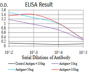
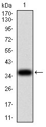
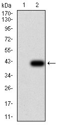
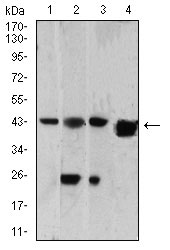
SH3GL1 Antibody
Purified Mouse Monoclonal Antibody
- SPECIFICATION
- CITATIONS
- PROTOCOLS
- BACKGROUND

Application
| WB, E |
|---|---|
| Primary Accession | Q99961 |
| Reactivity | Human, Rat |
| Host | Mouse |
| Clonality | Monoclonal |
| Clone Names | 4B4C2 |
| Isotype | IgG1 |
| Calculated MW | 41.5kDa |
| Description | This gene encodes a member of the endophilin family of Src homology 3 domain-containing proteins. The encoded protein is involved in endocytosis and may also play a role in the cell cycle. Overexpression of this gene may play a role in leukemogenesis, and the encoded protein has been implicated in acute myeloid leukemia as a fusion partner of the myeloid-lymphoid leukemia protein. Pseudogenes of this gene are located on the long arm of chromosomes 11 and 17. Alternatively spliced transcript variants encoding multiple isoforms have been observed for this gene. |
| Immunogen | Purified recombinant fragment of human SH3GL1 (AA: 12-119) expressed in E. Coli. |
| Formulation | Purified antibody in PBS with 0.05% sodium azide |
| Gene ID | 6455 |
|---|---|
| Other Names | Endophilin-A2, EEN fusion partner of MLL, Endophilin-2, Extra eleven-nineteen leukemia fusion gene protein, EEN, SH3 domain protein 2B, SH3 domain-containing GRB2-like protein 1, SH3GL1, CNSA1, SH3D2B |
| Dilution | WB~~1/500 - 1/2000 E~~1/10000 |
| Storage | Maintain refrigerated at 2-8°C for up to 6 months. For long term storage store at -20°C in small aliquots to prevent freeze-thaw cycles. |
| Precautions | SH3GL1 Antibody is for research use only and not for use in diagnostic or therapeutic procedures. |
| Name | SH3GL1 |
|---|---|
| Synonyms | CNSA1, SH3D2B |
| Function | Implicated in endocytosis. May recruit other proteins to membranes with high curvature (By similarity). |
| Cellular Location | Cytoplasm. Early endosome membrane; Peripheral membrane protein. Cell projection, podosome. Note=Associated with postsynaptic endosomes in hippocampal neurons. |
| Tissue Location | Ubiquitous. Higher expression in pancreas, placenta, prostate, testis and uterus |

Thousands of laboratories across the world have published research that depended on the performance of antibodies from Abcepta to advance their research. Check out links to articles that cite our products in major peer-reviewed journals, organized by research category.
info@abcepta.com, and receive a free "I Love Antibodies" mug.

Provided below are standard protocols that you may find useful for product applications.
References
1.J Exp Clin Cancer Res. 2012 Oct 11;31:85.2.Mol Med Rep. 2013 Oct;8(4):1111-7.
If you have used an Abcepta product and would like to share how it has performed, please click on the "Submit Review" button and provide the requested information. Our staff will examine and post your review and contact you if needed.
If you have any additional inquiries please email technical services at tech@abcepta.com.
Ordering Information
Other Products
Shipping Information
