


 Foundational characteristics of cancer include proliferation, angiogenesis, migration, evasion of apoptosis, and cellular immortality. Find key markers for these cellular processes and antibodies to detect them.
 The SUMOplot™ Analysis Program predicts and scores sumoylation sites in your protein. SUMOylation is a post-translational modification involved in various cellular processes, such as nuclear-cytosolic transport, transcriptional regulation, apoptosis, protein stability, response to stress, and progression through the cell cycle.
 The Autophagy Receptor Motif Plotter predicts and scores autophagy receptor binding sites in your protein. Identifying proteins connected to this pathway is critical to understanding the role of autophagy in physiological as well as pathological processes such as development, differentiation, neurodegenerative diseases, stress, infection, and cancer.
GNL3 Antibody
Purified Mouse Monoclonal Antibody
- SPECIFICATION
- CITATIONS
- PROTOCOLS
- BACKGROUND

Application
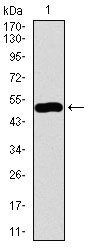
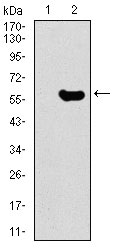
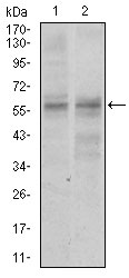
| WB, E |
|---|---|
| Primary Accession | Q9BVP2 |
| Reactivity | Human |
| Host | Mouse |
| Clonality | Monoclonal |
| Clone Names | 2C8D5 |
| Isotype | IgG1 |
| Calculated MW | 62kDa |
| Description | The protein encoded by this gene may interact with p53 and may be involved in tumorigenesis. The encoded protein also appears to be important for stem cell proliferation. This protein is found in both the nucleus and nucleolus. Three transcript variants encoding two different isoforms have been found for this gene. |
| Immunogen | Purified recombinant fragment of human GNL3 (AA: 1-226) expressed in E. Coli. |
| Formulation | Ascitic fluid containing 0.03% sodium azide. |
| Gene ID | 26354 |
|---|---|
| Other Names | Guanine nucleotide-binding protein-like 3, E2-induced gene 3 protein, Novel nucleolar protein 47, NNP47, Nucleolar GTP-binding protein 3, Nucleostemin, GNL3, E2IG3, NS |
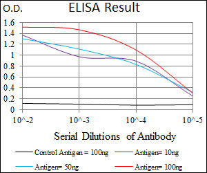
| Dilution | WB~~1/500 - 1/2000 E~~1/10000 |
| Storage | Maintain refrigerated at 2-8°C for up to 6 months. For long term storage store at -20°C in small aliquots to prevent freeze-thaw cycles. |
| Precautions | GNL3 Antibody is for research use only and not for use in diagnostic or therapeutic procedures. |
| Name | GNL3 |
|---|---|
| Synonyms | E2IG3, NS |
| Function | May be required to maintain the proliferative capacity of stem cells. Stabilizes MDM2 by preventing its ubiquitination, and hence proteasomal degradation (By similarity). |
| Cellular Location | Nucleus {ECO:0000250|UniProtKB:Q811S9}. Nucleus, nucleolus. Note=Shuttles between the nucleus and nucleolus. {ECO:0000250|UniProtKB:Q811S9} |
| Tissue Location | Increased levels in lung tissue in cancer patients. |

Thousands of laboratories across the world have published research that depended on the performance of antibodies from Abcepta to advance their research. Check out links to articles that cite our products in major peer-reviewed journals, organized by research category.
info@abcepta.com, and receive a free "I Love Antibodies" mug.

Provided below are standard protocols that you may find useful for product applications.
References
1.Oncogene. 2011 Apr 7;30(14):1716-26. 2.J Cell Biol. 2009 Jun 1;185(5):827-39.
If you have used an Abcepta product and would like to share how it has performed, please click on the "Submit Review" button and provide the requested information. Our staff will examine and post your review and contact you if needed.
If you have any additional inquiries please email technical services at tech@abcepta.com.
Ordering Information
Other Products
Shipping Information
