


 Foundational characteristics of cancer include proliferation, angiogenesis, migration, evasion of apoptosis, and cellular immortality. Find key markers for these cellular processes and antibodies to detect them.
 The SUMOplot™ Analysis Program predicts and scores sumoylation sites in your protein. SUMOylation is a post-translational modification involved in various cellular processes, such as nuclear-cytosolic transport, transcriptional regulation, apoptosis, protein stability, response to stress, and progression through the cell cycle.
 The Autophagy Receptor Motif Plotter predicts and scores autophagy receptor binding sites in your protein. Identifying proteins connected to this pathway is critical to understanding the role of autophagy in physiological as well as pathological processes such as development, differentiation, neurodegenerative diseases, stress, infection, and cancer.
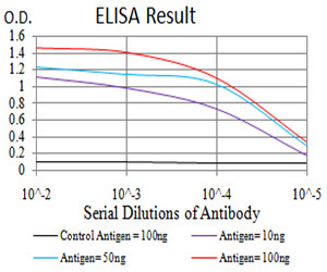
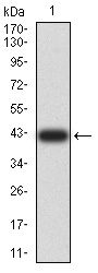
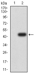
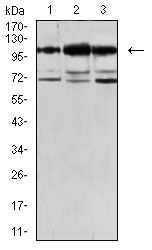
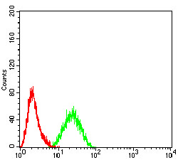
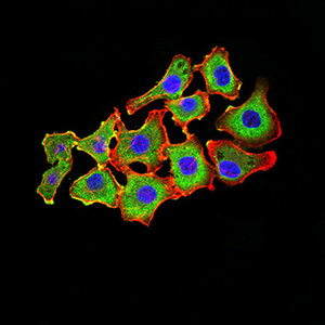
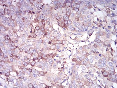
Mouse Monoclonal Antibody to CDH11
Purified Mouse Monoclonal Antibody
- SPECIFICATION
- CITATIONS
- PROTOCOLS
- BACKGROUND

Application
| WB, IHC, FC, ICC, E |
|---|---|
| Primary Accession | P55287 |
| Reactivity | Human |
| Host | Mouse |
| Clonality | Monoclonal |
| Clone Names | 2C12A6 |
| Isotype | Mouse IgG1 |
| Calculated MW | 88kDa |
| Description | This gene encodes a type II classical cadherin from the cadherin superfamily, integral membrane proteins that mediate calcium-dependent cell-cell adhesion. Mature cadherin proteins are composed of a large N-terminal extracellular domain, a single membrane-spanning domain, and a small, highly conserved C-terminal cytoplasmic domain. Type II (atypical) cadherins are defined based on their lack of a HAV cell adhesion recognition sequence specific to type I cadherins. Expression of this particular cadherin in osteoblastic cell lines, and its upregulation during differentiation, suggests a specific function in bone development and maintenance. ; |
| Immunogen | Purified recombinant fragment of human CDH11 (AA: 468-617) expressed in E. Coli. |
| Formulation | Purified antibody in PBS with 0.05% sodium azide |
| Application Note | ELISA: 1/10000 WB: 1/500 - 1/2000 IHC: 1/200 - 1/1000 ICC: 1/200 - 1/1000 FCM: 1/200 - 1/400 |
| Gene ID | 1009 |
|---|---|
| Other Names | OB; CAD11; CDHOB; OSF-4 |
| Dilution | WB~~1:1000 IHC~~1:100~500 FC~~1:10~50 ICC~~N/A E~~N/A |
| Storage | Maintain refrigerated at 2-8°C for up to 6 months. For long term storage store at -20°C in small aliquots to prevent freeze-thaw cycles. |
| Precautions | Mouse Monoclonal Antibody to CDH11 is for research use only and not for use in diagnostic or therapeutic procedures. |
| Name | CDH11 |
|---|---|
| Function | Cadherins are calcium-dependent cell adhesion proteins. They preferentially interact with themselves in a homophilic manner in connecting cells; cadherins may thus contribute to the sorting of heterogeneous cell types. Required for proper focal adhesion assembly (PubMed:33811546). Involved in the regulation of cell migration (PubMed:33811546). |
| Cellular Location | Cell membrane; Single-pass type I membrane protein |
| Tissue Location | Expressed mainly in brain but also found in other tissues. Expressed in neuroblasts. In the embryo from 67 to 72 days of gestation, detected at high levels in facial mesenchyme including the central palatal mesenchyme, dental mesenchyme, the eye and optic muscle, and the tongue (at protein level) (PubMed:33811546) |

Thousands of laboratories across the world have published research that depended on the performance of antibodies from Abcepta to advance their research. Check out links to articles that cite our products in major peer-reviewed journals, organized by research category.
info@abcepta.com, and receive a free "I Love Antibodies" mug.

Provided below are standard protocols that you may find useful for product applications.
References
1.Arthritis Rheumatol. 2014 Apr;66(4):1010-21. ; 2.Mol Cancer Res. 2012 Mar;10(3):293-304. ;
If you have used an Abcepta product and would like to share how it has performed, please click on the "Submit Review" button and provide the requested information. Our staff will examine and post your review and contact you if needed.
If you have any additional inquiries please email technical services at tech@abcepta.com.
Ordering Information
Other Products
Shipping Information
