


 Foundational characteristics of cancer include proliferation, angiogenesis, migration, evasion of apoptosis, and cellular immortality. Find key markers for these cellular processes and antibodies to detect them.
 The SUMOplot™ Analysis Program predicts and scores sumoylation sites in your protein. SUMOylation is a post-translational modification involved in various cellular processes, such as nuclear-cytosolic transport, transcriptional regulation, apoptosis, protein stability, response to stress, and progression through the cell cycle.
 The Autophagy Receptor Motif Plotter predicts and scores autophagy receptor binding sites in your protein. Identifying proteins connected to this pathway is critical to understanding the role of autophagy in physiological as well as pathological processes such as development, differentiation, neurodegenerative diseases, stress, infection, and cancer.
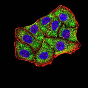
Mouse Monoclonal Antibody to BNIP3
Purified Mouse Monoclonal Antibody
- SPECIFICATION
- CITATIONS
- PROTOCOLS
- BACKGROUND

Application
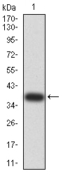
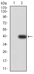
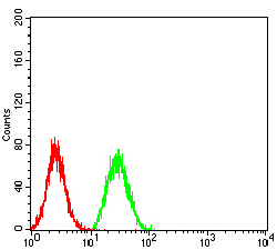
| WB, FC, ICC, E |
|---|---|
| Primary Accession | Q12983 |
| Reactivity | Human |
| Host | Mouse |
| Clonality | Monoclonal |
| Clone Names | 4E11F4 |
| Isotype | Mouse IgG1 |
| Calculated MW | 21.5kDa |
| Description | This gene is encodes a mitochondrial protein that contains a BH3 domain and acts as a pro-apoptotic factor. The encoded protein interacts with anti-apoptotic proteins, including the E1B 19 kDa protein and Bcl2. This gene is silenced in tumors by DNA methylation.; |
| Immunogen | Purified recombinant fragment of human BNIP3 (AA: 50-155) expressed in E. Coli. |
| Formulation | Purified antibody in PBS with 0.05% sodium azide |
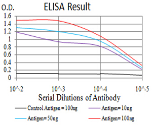
| Application Note | ELISA: 1/10000 WB: 1/500 - 1/2000 ICC: 1/200 - 1/1000 FCM: 1/200 - 1/400 |
| Gene ID | 664 |
|---|---|
| Other Names | NIP3 |
| Dilution | WB~~1:1000 FC~~1:10~50 ICC~~N/A E~~N/A |
| Storage | Maintain refrigerated at 2-8°C for up to 6 months. For long term storage store at -20°C in small aliquots to prevent freeze-thaw cycles. |
| Precautions | Mouse Monoclonal Antibody to BNIP3 is for research use only and not for use in diagnostic or therapeutic procedures. |
| Name | BNIP3 (HGNC:1084) |
|---|---|
| Synonyms | NIP3 |
| Function | Apoptosis-inducing protein that can overcome BCL2 suppression. May play a role in repartitioning calcium between the two major intracellular calcium stores in association with BCL2. Involved in mitochondrial quality control via its interaction with SPATA18/MIEAP: in response to mitochondrial damage, participates in mitochondrial protein catabolic process (also named MALM) leading to the degradation of damaged proteins inside mitochondria. The physical interaction of SPATA18/MIEAP, BNIP3 and BNIP3L/NIX at the mitochondrial outer membrane regulates the opening of a pore in the mitochondrial double membrane in order to mediate the translocation of lysosomal proteins from the cytoplasm to the mitochondrial matrix. Plays an important role in the calprotectin (S100A8/A9)-induced cell death pathway. |
| Cellular Location | Mitochondrion. Mitochondrion outer membrane; Single-pass membrane protein. Note=Coexpression with the EIB 19-kDa protein results in a shift in NIP3 localization pattern to the nuclear envelope. Colocalizes with ACAA2 in the mitochondria. Colocalizes with SPATA18 at the mitochondrion outer membrane |

Thousands of laboratories across the world have published research that depended on the performance of antibodies from Abcepta to advance their research. Check out links to articles that cite our products in major peer-reviewed journals, organized by research category.
info@abcepta.com, and receive a free "I Love Antibodies" mug.

Provided below are standard protocols that you may find useful for product applications.
References
1.Tumour Biol. 2015 Jun;36(6):4731-40. ; 2.PLoS One. 2014 May 13;9(5):e96733. ;
If you have used an Abcepta product and would like to share how it has performed, please click on the "Submit Review" button and provide the requested information. Our staff will examine and post your review and contact you if needed.
If you have any additional inquiries please email technical services at tech@abcepta.com.
Ordering Information
Other Products
Shipping Information
